Melanoma
Esther Oliva, MD
Key Facts
Clinical Issues
Age range: 20-50 years
Macroscopic Features
Black cut surface (occasionally)
Microscopic Pathology
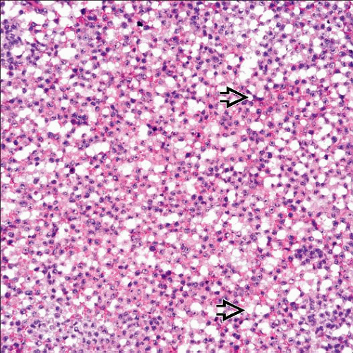
Diffuse, nested, pseudopapillary (rare) growth
Follicle-like spaces may be seen
Associated mature cystic teratoma (most often)
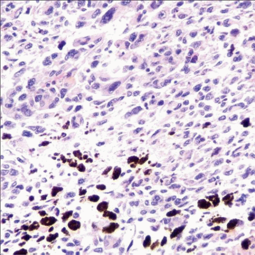
Epithelioid cells (most common), spindle or small
Signet ring-like or giant cells rare
Melanin pigment and intranuclear pseudoinclusions
Ancillary Tests
HMB-45, S100, Melan-A, tyrosinase, MITF, SOX10 (+)
Top Differential Diagnoses
Metastatic malignant melanoma
Poorly differentiated or undifferentiated carcinoma
Steroid cell tumor
TERMINOLOGY
Definitions
Malignant neoplasm showing melanocytic differentiation, most often arising in a background of mature cystic teratoma
ETIOLOGY/PATHOGENESIS
Malignant Transformation
From melanocytes present in ectodermal derivatives of mature cystic teratoma
CLINICAL ISSUES
Epidemiology
Incidence
Rare
Age
Most commonly occurs from age 20-50 years
Stay updated, free articles. Join our Telegram channel

Full access? Get Clinical Tree


