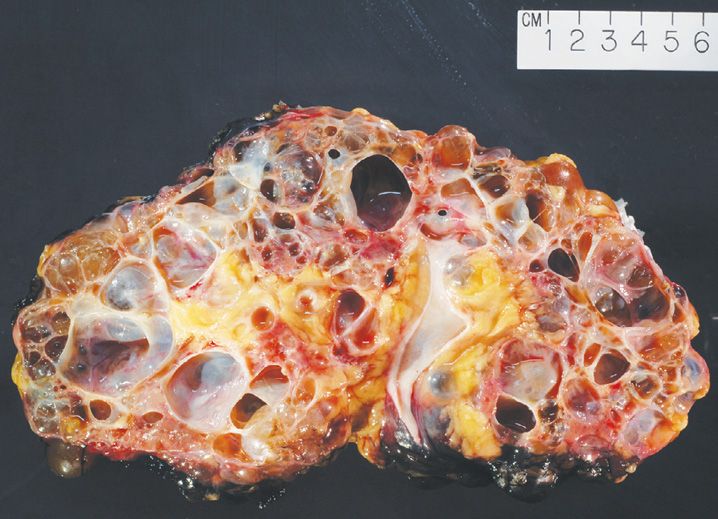
FIGURE 19-1
(A) Autosomal-dominant polycystic kidney disease
(B) Autosomal-recessive polycystic kidney disease
(C) Cystic renal dysplasia
(D) Medullary sponge kidney
(E) Simple renal cysts
4. A nephrectomy specimen from a 40-year-old man is shown in Figure 19-2. Both the kidneys have a similar appearance. Which of the following statements does not apply to this condition?
(A) Adult form shows an autosomal-recessive pattern of inheritance
(B) Cysts initially involve only portion of a nephron
(C) 40% patients have polycystic liver disease
(D) Mutations in PKD1 occur in 85% of patients
(E) Renal function is maintained until 4th or 5th decade of life
5. Which of the following mechanisms of glomerular injury causes a diffuse linear pattern of staining by immunofluorescence?
(A) Antibodies against antigen complex located on the basal surface of visceral epithelial cells
(B) Antibodies against exogenous/endogenous antigens not present in the glomerulus (“planted antigens”)
(C) Antibodies directed against circulating tumor antigens
(D) Antibodies directed against circulating Hepatitis C virus antigen
(E) Antibodies against NC1 domain of collagen type IV in glomerular basement membrane
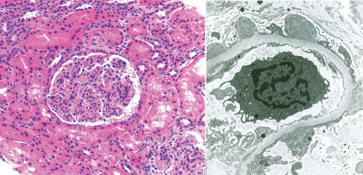
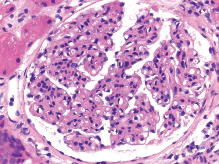
6. A 10-year-old boy with history of fever and impetigo presents with abrupt onset oliguria and hematuria. A renal biopsy and the corresponding electron microscopic findings are shown in Figure 19-3. What is the best diagnosis?
(A) Acute poststreptococcal glomerulonephritis
(B) Goodpasture syndrome
(C) Idiopathic rapidly progressing glomerulonephritis
(D) Membranous glomerulopathy
(E) Minimal change disease
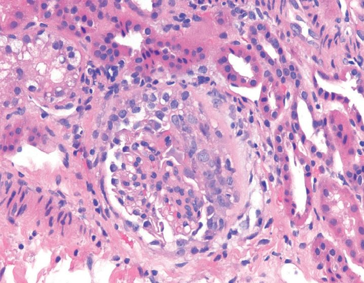
7. A 45-year-old man presents with rapid and progressive deterioration of renal function and severe oliguria. His renal biopsy shows this finding in most of the glomeruli (see Figure 19-4). All the following statements are associated with this condition, except
(A) Electron microscopy shows no changes
(B) Fatal, if the condition is left untreated
(C) Greater than 90% of patients with type III disease have circulating antineutrophil cytoplasmic antibodies
(D) Type I form of this disease is associated with anti-glomerular basement membrane antibodies
(E) Type II form of this disease is associated with immune complex deposits
8. Which of the following glomerular diseases is the most common cause of nephrotic syndrome in children?
(A) Focal segmental glomerulosclerosis
(B) IgA nephropathy
(C) Membranoproliferative glomerulonephritis
(D) Membranous glomerulopathy
(E) Minimal change disease
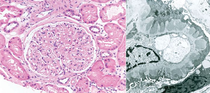
9. A renal biopsy and electron microscopic findings from a 53-year-old man with history of nephrotic syndrome is shown in Figure 19-5. Which of the following conditions is the most common cause of this pattern of renal injury?
(A) Drugs
(B) Idiopathic
(C) Infections
(D) Malignancy
(E) Systemic lupus erythematosus
10. A 2-year-old boy following routine prophylactic immunization is found to have highly selective proteinuria. Based on the clinical suspicion of minimal change disease, steroid therapy is instituted, and his condition rapidly returns to normal. Which of the following statements is least likely to be associated with this condition?
(A) Excellent long-term prognosis
(B) Hypercellular glomeruli
(C) Intracytoplasmic lipid and protein droplets in proximal tubules
(D) Normal blood pressure
(E) Uniform and diffuse effacement of foot processes on electron microscopy
11. Which of the following patterns of glomerular injury is common to patients with HIV infection, heroin addicts, and sickle cell disease?
(A) Focal segmental glomerulosclerosis
(B) Membranoproliferative glomerulonephritis
(C) Membranous glomerulopathy
(D) Minimal change disease
(E) Rapidly progressive glomerulonephritis
12. Which of the following forms of glomerular diseases is the most common cause of nephrotic syndrome in adults in United States?
(A) Focal segmental glomerulosclerosis
(B) Membranoproliferative glomerulonephritis
(C) Membranous glomerulopathy
(D) Minimal change disease
(E) Rapidly progressive glomerulonephritis
13. IgA nephropathy is characterized by all of the following features, except
(A) Frequent recurrence in transplanted kidneys
(B) Increased association with celiac disease and liver disease
(C) Most common cause of glomerulonephritis worldwide
(D) Monoclonal deposits of IgA in mesangial region
(E) Recurrent gross and/or microscopic hematuria
14. A 15-year-old teenager with history of gross hematuria and vision problems is found to have nerve deafness and dislocation of the left ocular lens. Electron microscopy study shows irregular foci of thickening and thinning of the glomerular basement membrane with a “basket-weave” appearance of the lamina densa. Which of the following choices best describes this renal condition?
(A) Alport syndrome
(B) IgA nephropathy
(C) Minimal change disease
(D) Poststreptococcal glomerulonephritis
(E) Thin basement membrane disease
15. Which of the following conditions is least likely to progress towards chronic glomerulonephritis?
(A) Focal segmental glomerulosclerosis
(B) Membranoproliferative glomerulonephritis
(C) Membranous glomerulopathy
(D) Poststreptococcal glomerulonephritis
(E) Rapidly progressive glomerulonephritis
16. A biopsy from a patient with active lupus nephritis is shown in Figure 19-6. Based on the pattern of disease shown here, in which of the following portions of the nephron are the immune complexes typically deposited?
(A) Intramembranous
(B) Mesangial
(C) Subendothelial
(D) Subepithelial
(E) Tubular
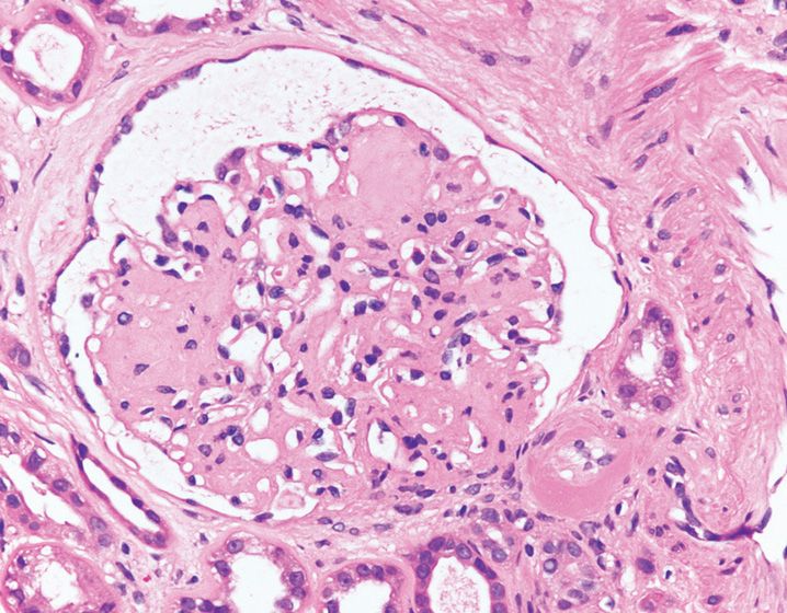
17. A renal biopsy obtained from a patient with history of microalbuminuria is shown in Figure 19-7. What is the best diagnosis?
Stay updated, free articles. Join our Telegram channel

Full access? Get Clinical Tree


