Mediastinal Seminoma
Key Facts
Terminology
Thymic seminoma
Etiology/Pathogenesis
Probably misplaced germ cells in anterior mediastinum
Clinical Issues
Incidence
May account for approximately 37% of all germ cell tumors
Majority of tumors occur in young adults between ages of 20 and 30 years
Almost exclusive occurrence in men
Symptoms
Gynecomastia
Superior vena cava syndrome
Chest pain
Macroscopic Features
Large tumors
Cystic tumor can occur in < 10% of cases
Top Differential Diagnoses
Carcinoma
Lymphoma
Melanoma
Thymoma
Metastatic seminoma from testicular origin
Diagnostic Checklist
Sheets of neoplastic cells
Inflammatory component (lymphocytes)
Cells with clear or eosinophilic cytoplasm
PLAP positive immunohistochemical study
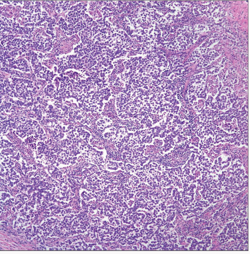 Low-power view of a thymic seminoma is shown with sheets of neoplastic cells with little intervening stroma and scattered inflammatory cells. |
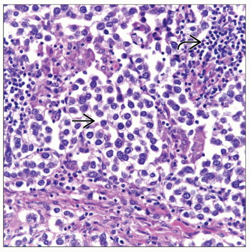 High-power view of a thymic seminoma displays sheets of neoplastic cells  with a discrete inflammatory reaction composed of lymphocytes with a discrete inflammatory reaction composed of lymphocytes  . . |
TERMINOLOGY
Synonyms
Thymic seminoma, germinoma
Definitions
Malignant germ cell tumor
ETIOLOGY/PATHOGENESIS
Etiology
Although etiology of these tumors is unknown, some theories have stated that misplaced germ cells in anterior mediastinum may be origin of these tumors
CLINICAL ISSUES
Epidemiology
Incidence
Difficult to determine exact incidence
2nd most common germ cell tumor after teratomas
May account for approximately 37% of all germ cell tumors
Age
Majority of tumors occur in young adults between ages of 20 and 30 years
Highly unusual in patients < 12 years old
Minority of cases in adult patients > 60 years old
Gender
Almost exclusive occurrence in males
Only a few cases reported in females
Site
Anterior mediastinal tumor
Rarely in posterior mediastinum
Presentation
Shortness of breath
Cough
Chest pain
Hemoptysis
Superior vena cava syndrome
Gynecomastia
Pulmonic stenosis
Ventricular septal defect
Congenital absence of thoracic hemivertebrae
In some cases, patients may be asymptomatic
Treatment
Chemotherapy
Radiation therapy
Prognosis
Depends on clinical staging
May be better in younger than in older patients
IMAGE FINDINGS
General Features
Large, bulky tumors
Well marginated
May extend to both sides of the midline
CT Findings
Homogeneous attenuation equal to soft tissue
MACROSCOPIC FEATURES
General Features
Large tumors
Slightly lobulated, firm and tan
Cystic tumor can occur in < 10% of cases
Size
Vary in size from a few cm to > 15 cm in greatest dimension
MICROSCOPIC PATHOLOGY
Histologic Features
Sheets or discrete nesting pattern
Medium-sized cells
Clear or lightly eosinophilic cytoplasm
Inflammatory infiltrate (lymphocytes)
Predominant Pattern/Injury Type
Sheets
Predominant Cell/Compartment Type
Germ, seminomatous
DIFFERENTIAL DIAGNOSIS
Carcinoma
Displays lobulation and more cellular pleomorphism
Positive reaction for PLAP unusual
Lymphoma
May show extensive areas of fibrosis
Shows negative staining for CK-LMW-NOS and PLAP
Melanoma
Shows positive staining for S100 protein and mart-1; negative for keratin and PLAP
Thymoma
Shows classical features of lobulation and mixed biphasic cellular components
Metastatic Seminoma from Testicular Origin
Rarely metastasizes as bulky anterior mediastinal tumor
May metastasize to mediastinal lymph nodes
Identical histopathological and immunohistochemical features for both tumors (mediastinal and testicular)
DIAGNOSTIC CHECKLIST
Clinically Relevant Pathologic Features
Age distribution
Pathologic Interpretation Pearls
Sheets of neoplastic cells
Inflammatory component (lymphocytes)
Cells with clear or eosinophilic cytoplasm
PLAP positive immunohistochemical study
SELECTED REFERENCES
1. Giannis M et al: Cisplatin-based chemotherapy for advanced seminoma: report of 52 cases treated in two institutions. J Cancer Res Clin Oncol. 135(11):1495-500, 2009
2. Sung MT et al: Primary mediastinal seminoma: a comprehensive assessment integrated with histology, immunohistochemistry, and fluorescence in situ hybridization for chromosome 12p abnormalities in 23 cases. Am J Surg Pathol. 32(1):146-55, 2008
3. Malagón HD et al: Germ cell tumors with sarcomatous components: a clinicopathologic and immunohistochemical study of 46 cases. Am J Surg Pathol. 31(9):1356-62, 2007
4. Bedano PM et al: Metachronous intracranial germinoma in a patient with a previous primary mediastinal seminoma. J Clin Oncol. 24(15):2386-7, 2006
5. Miyawaki M et al: [High-dose chemotherapy with peripheral blood stem cell auto transplantation for an intractable case of mediastinal seminoma.] Gan To Kagaku Ryoho. 33(6):845-8, 2006
Stay updated, free articles. Join our Telegram channel

Full access? Get Clinical Tree


