Malignant Rhabdoid Tumor
Satish K. Tickoo, MD
Victor E. Reuter, MD
Key Facts
Terminology
Malignant rhabdoid tumor of kidney (RTK)
Highly malignant pediatric renal tumor with very poor prognosis and genetic abnormalities of hSNF5/INI1 tumor suppressor gene on chromosome 22
Etiology/Pathogenesis
Biallelic inactivation of gene, located at 22q11.2; consistent feature of RTK
Clinical Issues
Mean age of presentation: Around 1 year old
Predominantly affects younger children; 80% < 2 years old, 60% < 1 year old
Overwhelming majority of stage IV renal tumors in 1st 7 months of life are RTK
High tumor stage at presentation
Macroscopic Features
Well-circumscribed and unencapsulated
Foci of hemorrhage and necrosis
Microscopic Pathology
Sheets of monotonous, loosely cohesive, large ovoid to polygonal cells, with high nuclear grade
Characteristic cytologic features: Vesicular chromatin, prominent eosinophilic nucleoli, and intracytoplasmic hyaline, pink inclusion, at least in some cells
Ancillary Tests
SNF5(INI1) negative immunostaining considered specific
Reliable surrogate marker of hSNF5/INI1 gene deletion or inactivating mutations
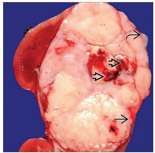 RTK is seen with necrosis  , irregular invasive borders , irregular invasive borders  , and extension beyond renal parenchyma , and extension beyond renal parenchyma  . Necrosis may be more extensive, and some tumors may be relatively small due to early dissemination. . Necrosis may be more extensive, and some tumors may be relatively small due to early dissemination. |
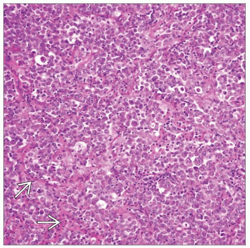 Sheets of loosely cohesive tumor cells with large nuclei and abundant eosinophilic cytoplasm are typical of RTK. A delicate network of fibrovascular septae  may also be appreciated. may also be appreciated. |
TERMINOLOGY
Abbreviations
Malignant rhabdoid tumor of kidney (RTK)
Definitions
Highly malignant pediatric renal tumor with very poor prognosis and genetic abnormalities of hSNF5/INI1 tumor suppressor gene on chromosome 22
ETIOLOGY/PATHOGENESIS
HSNF5/INI1 Tumor Suppressor Gene
Biallelic inactivation of gene, located at 22q11.2; consistent feature of RTK
Usually associated with deletion of 1 copy with mutation in remaining copy
Gene believed to be important for chromatin remodeling
CLINICAL ISSUES
Epidemiology
Incidence
Comprises approximately 2% of pediatric renal tumors
Age
Mean age of presentation around 1 year
Predominantly affects younger children; 80% < 2 years old, 60% < 1 year old
Virtually nonexistent after 5 years of age
Overwhelming majority of stage IV renal tumors in 1st 7 months of life are RTK
Gender
M:F = 1.5:1
Site
Originally described in kidney; similar tumors later recognized in extrarenal sites, including
Central nervous system (atypical teratoid/rhabdoid tumor) and soft tissue
Presentation
Abdominal mass; most common mode of presentation
Stay updated, free articles. Join our Telegram channel

Full access? Get Clinical Tree


