Lymphoepithelioma-like Carcinoma
Key Facts
Terminology
Lymphoepithelioma-like carcinoma (LELC)
Malignant epithelial neoplasm with prominent inflammatory component
Etiology/Pathogenesis
Appears not to be related to tobacco smoke
Closely associated with Epstein-Barr virus (EBV) infection
More common in people of Asian background though also described in Caucasians
Microscopic Pathology
Inflammatory
Epithelial
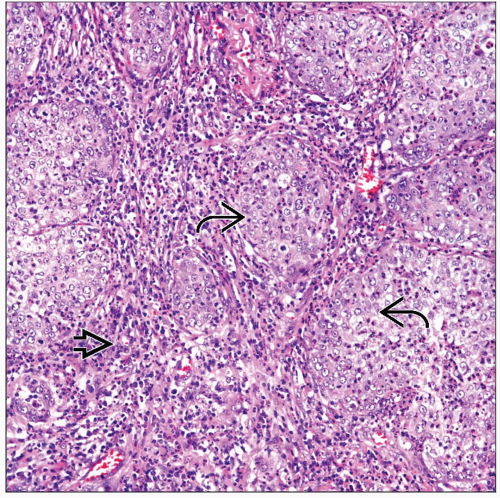
Malignant epithelial component embedded in inflammatory background composed predominantly of lymphocytes and plasma cells
Top Differential Diagnoses
Metastatic nasopharyngeal carcinoma
Identical histological features to counterpart in head and neck
Clinical history &/or physical examination play important role in determining primary site
Large cell carcinoma
Presence of prominent inflammatory component should raise suspicion of LELC
Diagnostic Checklist
Histological features similar to nasopharyngeal carcinoma (Schmincke tumor)
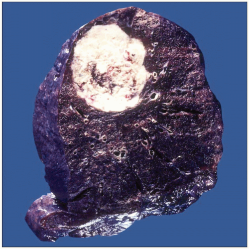
 Gross photograph of a lymphoepithelial-like carcinoma shows a well-circumscribed intrapulmonary mass with focal central necrosis. The tumor has a peripheral subpleural location. |
TERMINOLOGY
Abbreviations
Lymphoepithelioma-like carcinoma (LELC)
Definitions
Malignant epithelial neoplasm with prominent inflammatory component
ETIOLOGY/PATHOGENESIS
Environmental Exposure
Appears not to be related to tobacco smoke
Infectious Agents
Closely associated with Epstein-Barr virus (EBV) infection
More common in people of Asian background though also described in Caucasians
CLINICAL ISSUES
Presentation
Cough
Hemoptysis
Difficulty breathing
Chest pain
Weight loss
Treatment
Surgical approaches
Lobectomy
Adjuvant therapy
Chemotherapy or radiation therapy may be used depending on stage
Prognosis
Depends on stage at time of diagnosis
MACROSCOPIC FEATURES
General Features
Tumor may be in central or peripheral location
Essentially indistinguishable from other non-small cell carcinomas
Necrosis is common finding
Size
Tumors may vary in size and may be > 10 cm
MICROSCOPIC PATHOLOGY
Histologic Features
Large epithelial cells with scant cytoplasm and vesicular nuclei
Lobulated pattern of growth
Cells with round to oval nuclei and prominent nucleoli
Nuclear atypia and increased mitotic activity
Prominent inflammatory background
Lymphocytes and plasma cells easily identified
EBER studies are positive in tumor cells
Predominant Pattern/Injury Type
Mixed
Inflammatory
Predominant Cell/Compartment Type
Epithelial
DIFFERENTIAL DIAGNOSIS
Metastatic Nasopharyngeal Carcinoma
Identical histological features to counterpart in head and neck
Clinical history &/or physical examination play important role in determining primary site
Immunohistochemical features may be similar
Large Cell Carcinoma
Presence of prominent inflammatory component should raise suspicion of LELC
Will display sheets of large neoplastic cells with prominent nucleoli
Extensive areas of necrosis &/or hemorrhage commonly seen
Stay updated, free articles. Join our Telegram channel

Full access? Get Clinical Tree


