Low-Grade Fibromyxoid Sarcoma (Evans Tumor)
Khin Thway, BSc, MBBS, FRCPath
Key Facts
Terminology
Malignant fibroblastic neoplasm composed of bland spindle cells in collagenous matrix, often with prominent collagenous nodules
Characterized by specific translocations producing fusion oncogenes
Clinical Issues
Adults (typically in 4th decade)
Mainly deep-seated, but can also occur superficially in dermis and subcutis
Proximal extremities (especially lower limbs) and trunk; rarely other sites
Recurrence rates of up to 21%
Recurrence lower in superficial cases
Metastatic rate of approximately 30% in genetically confirmed cases
Microscopic Pathology
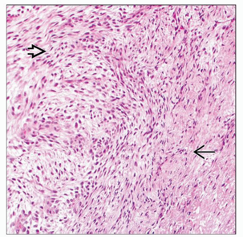
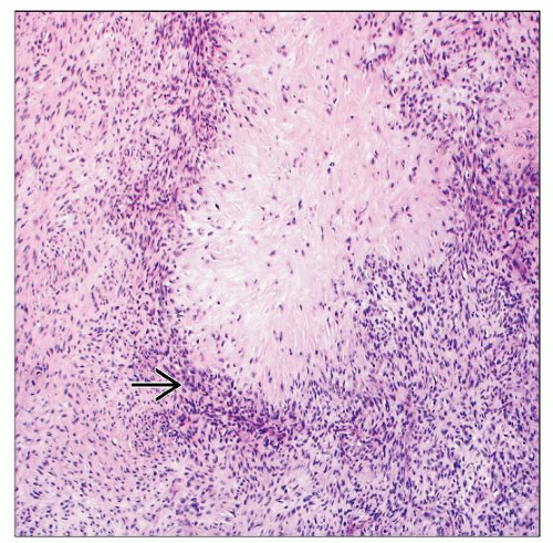
Whorled distributions of bland fibroblasts
Collagenous or myxoid matrix, often in distinct zones
Owing to bland morphology, tumors often mistaken for variety of benign or low-grade neoplasms
Ancillary Tests
MUC4 immunohistochemistry is sensitive and specific
CD34 positivity in some cases
Occasional EMA and claudin-1 positivity, which can make distinction from perineurioma difficult
Balanced translocations
t(7;16)(q32-34;p11) FUS, CREB3L2
t(11;16)(p11;p11) FUS, CREB3L1
TERMINOLOGY
Abbreviations
Low-grade fibromyxoid sarcoma (LGFMS)
Synonyms
Fibrosarcoma, fibromyxoid type; hyalinizing spindle cell tumor with giant rosettes
Definitions
Malignant fibroblastic neoplasm composed of bland spindle cells in collagenous and myxoid matrix, often with prominent collagenous nodules
Characterized by specific translocations producing fusion oncogenes
Despite histologically low-grade morphology, up to 30% can metastasize
ETIOLOGY/PATHOGENESIS
Characteristic Translocations
Produce chimeric fusion genes
Cellular origin still unknown, but mesenchymal neoplasm with fibroblastic-like cells
CLINICAL ISSUES
Epidemiology
Incidence
True incidence unknown
Tumor probably underreported in literature due to its morphologic resemblance to other benign and malignant tumors
Age
Adults (typically in 4th decade), but wide age distribution
Significant proportion in patients < 18 years old
Gender
M > F
Site
Mainly deep-seated, but a smaller proportion occurs in dermis and subcutis
Proximal extremities (especially lower limbs) and trunk; rarely other sites including viscera
Superficial lesions have higher incidence in childhood
Presentation
Painless mass
Many are of long duration
Treatment
Surgical approaches
Wide excision
Long-term follow-up is mandatory, in view of potential for late metastases
Prognosis
Recurrence rates of up to 21%
Recurrence is lower in superficial cases
Metastatic rate of approximately 30% in genetically confirmed cases
> 80% of metastases appeared after 9 years
No reported metastases in superficial tumors
MACROSCOPIC FEATURES
General Features
Well-defined mass
White cut surface, often with glistening myxoid areas
Sometimes cystic foci, but necrosis rare
MICROSCOPIC PATHOLOGY
Histologic Features
Lobulated and partially circumscribed, but frequent microscopic infiltration into adjacent soft tissue
Stay updated, free articles. Join our Telegram channel

Full access? Get Clinical Tree


