13 Liver Diseases
Anatomy of the Liver
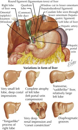

Basic Gross Anatomy


• Hepatoduodenal ligament: peritoneal fold surrounding portal triad (hepatic artery proper, portal vein, bile duct), right edge of lesser omentum
• Omental foramen (of Winslow): posterior to hepatoduodenal ligament, opens into lesser (peritoneal) sac
Divisions
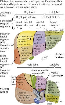
• Right and left hepatic lobes: divided by a plane extending from cystic fossa (anteroinferior) through inferior vena cava (superoposterior)

• Each hemiliver contains its own hepatic artery branch, portal blood supply, venous drainage, and bile duct.


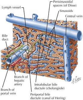
Portal Triads and Bile Duct System
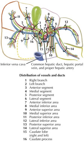
• Portal triads—hepatic artery, bile duct, and portal vein branches—and lymphatics seen in characteristic relationships from microscopic (lobular) to macroscopic (lobar) levels
• Popular functional concepts of liver parenchyma include classic lobules and liver acini organized around vessels.
• Interlobular hepatic artery branches travel alongside portal veins in septa, providing smaller branches to ducts and parenchyma (hepatocytes) of lobules.