Leiomyosarcoma
Key Facts
Clinical Issues
Incidence
Represents < 1% of all malignant neoplasms of lung
Described in any age group from newborns to adults
Symptoms
Cough
Hemoptysis
Chest pain
Dyspnea
Weight loss
Asymptomatic
Macroscopic Features
Endobronchial or peripheral
Areas of hemorrhage and necrosis may be present
Tumors vary in size from 1 to > 10 cm in diameter
Top Differential Diagnoses
Hemangiopericytoma
Usually does not display marked cellular pleomorphisms
Shows negative staining for muscle markers
Monophasic synovial sarcoma
Usually shows a more cohesive spindle cell proliferation
Generally shows negative results with muscle markers
Usually shows positive staining for keratin, EMA/MUC1, and Bcl-2
Solitary fibrous tumor
Usually shows positive staining for CD34 and Bcl-2
Displays negative staining for muscle markers
TERMINOLOGY
Abbreviations
Pulmonary leiomyosarcoma (LMS)
Definitions
Malignant mesenchymal neoplasm of smooth muscle origin
CLINICAL ISSUES
Epidemiology
Incidence
Represents < 1% of all malignant neoplasms of lung
Age
Has been described in any age group from newborns to adult patients
Gender
No predilection for any gender
Presentation
Cough
Hemoptysis
Chest pain
Shortness of breath
Weight loss
Asymptomatic
Treatment
Surgical approaches
Lobectomy or pneumonectomy
Adjuvant therapy
Chemo-radiation may be used in some cases
Prognosis
Depends on tumor differentiation and clinical staging
Widespread metastatic disease can occur
Some patients may follow aggressive course with death in < 24 months
MACROSCOPIC FEATURES
General Features
Well-circumscribed tumors
Endobronchial or peripheral
Cut surface is white and firm
Areas of hemorrhage and necrosis may be present
Size
Tumor varies in size from 1 cm to > 10 cm in diameter
MICROSCOPIC PATHOLOGY
Histologic Features
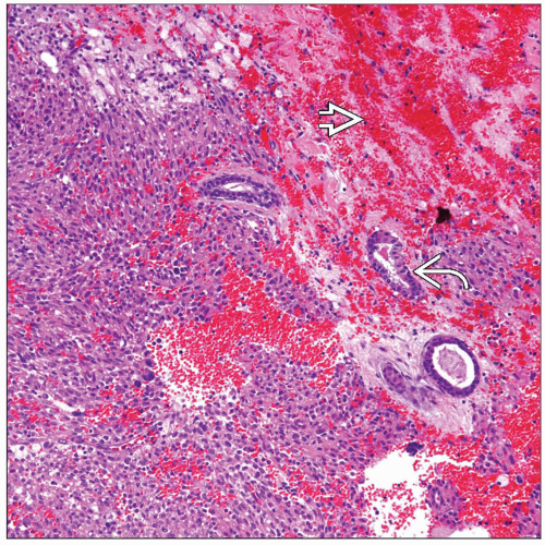
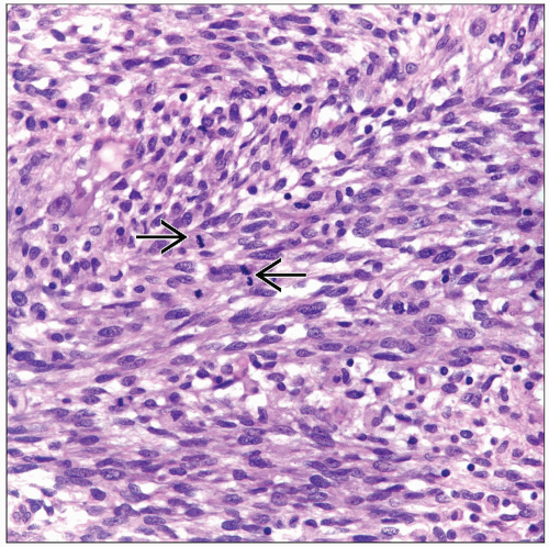
Spindle cell proliferation of elongated cells with cigar-shaped nuclei
Predominant Pattern/Injury Type
Stay updated, free articles. Join our Telegram channel

Full access? Get Clinical Tree


