Keloid and Cellular Scar
David S. Cassarino, MD, PhD
Key Facts
Terminology
Scar with prominent thickened and eosinophilic bundles of collagen
Clinical Issues
Persistence and recurrence are common, but no risk of malignancy
Scar that grows beyond original wound
Often erythematous, pruritic lesions with predilection for earlobe in black patients
Microscopic Pathology
Dense proliferation of thickened, hyalinized collagen bundles in dermis
Decreased vessels compared to conventional and hypertrophic scars
Increased fibroblasts, lymphocytes, and mast cells are usually present
Top Differential Diagnoses
Hypertrophic scar
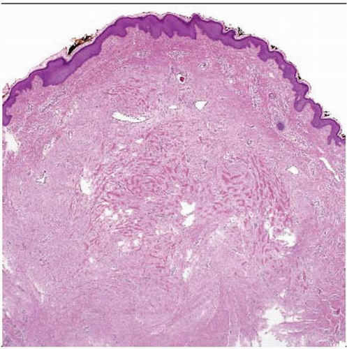 Low-power examination shows a polypoid skin lesion with dense dermal collagen. Note the mild epidermal hyperplasia. Commonly the epidermis overlying a keloid scar is atrophied. |
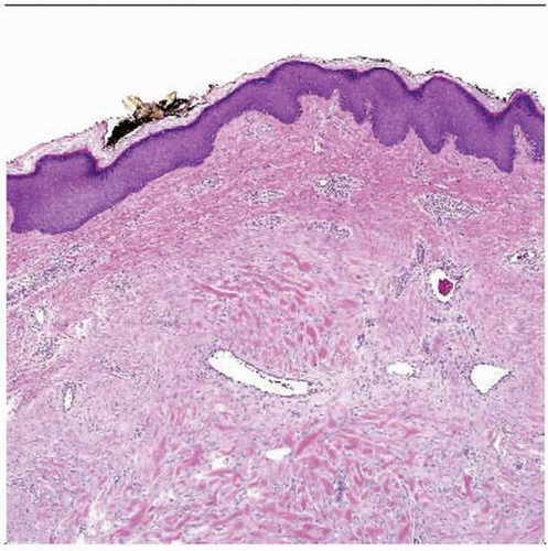 Hematoxylin & eosin shows the superficial aspect of the lesion. Note the dermal scarring with telangiectasia and prominent hyalinized collagen bundles. |
TERMINOLOGY
Synonyms
Scar with keloidal collagen
Definitions
Scar with prominent thickened and eosinophilic bundles of collagen extending beyond original wound
ETIOLOGY/PATHOGENESIS
Unknown, Possibly Genetic
Fibroblasts from keloids show decreased apoptosis
Many cytokines implicated in stimulating fibroblasts, including TGF-β1 and IL-15
CLINICAL ISSUES
Epidemiology
Age
Most common in patients < 30 years
Ethnicity
More common in black patients; least common in Caucasians
Site
Earlobe is most common site
Typically follows ear piercing or other trauma by a few months


