12 ITU
Shock
Case history (1)
A 60-year-old bank manager presents with crushing central chest pain. This started 45 minutes ago and has remained constant ever since.
On examination he is pale and clammy. His pulse is 100/min, poor volume and BP 90/60.
Diagnosis.
Shock – cardiogenic; ECG shows evidence of anterior myocardial infarction.
Case history (2)
A 50-year-old man who works in a bar has been brought to hospital vomiting a large amount of blood. He gave no history of upper abdominal pain but did admit to drinking a bottle of whiskey a day and occasional beers as well.
He also admits to a previous hospital admission with abdominal swelling which was due to alcoholic liver disease. He was told that his liver disease was bad and that he must stop drinking and take propranolol regularly as he had varices on endoscopy. He had not stopped drinking, nor was he taking his propranolol.
On examination he is sweating profusely with visible shaking of his extremeties. His pulse rate is 120/min with a blood pressure of 90/60.
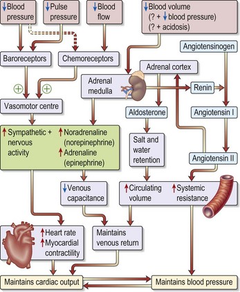
Physiology (Fig. 12.1)
Shock is ‘inadequate tissue oxygenation’ where there is failure of the circulatory system due to:
• Failure of the heart to maintain an adequate cardiac output, e.g. myocardial infarction
• Reduction in the volume of blood within the circulation, e.g. haemorrhage
• Loss of vascular tone within the circulatory system, e.g. septicaemia
Causes of shock
• Cardiogenic: myocardial infarction, tamponade, aortic dissection, pulmonary embolism
• Sepsis: infective causes, e.g. pneumonia, urinary tract infection or meningococcal septicaemia or ‘systemic inflammation’, resulting from trauma, post-surgery or anaphylaxis
Is the shock state in Case 1 due to myocardial infarction, an obstructed circulation or other shock-inducing factors?
A history in a shocked patient of chest pain associated with ECG evidence of a myocardial infarction makes the diagnosis in this case. However, be careful because ECG changes with tachyarrhythmias and some ST segment changes which look characteristic of myocardial ischaemia can also occur in other types of shock.
Is the shock state in Case 2 due to fluid loss? If so, what fluid and from where?
In this case the blood loss is obvious, i.e. haematemesis. Try and estimate from the patient how much was lost. In other cases blood loss might be concealed, e.g. pancreatitis.
Clinical examination
In assessing the shocked patient the following indices should be monitored:
Case 1 – has a problem with the circulatory pump and probably needs no extra intravascular fluid administration.
Case 2 – the haematemesis patient – has lost circulating volume with a low central venous pressure. His problems are with the plumbing circuit not the heart pump but his condition might be compounded by coagulation and biochemical disturbances consequent upon hepatic insufficiency.
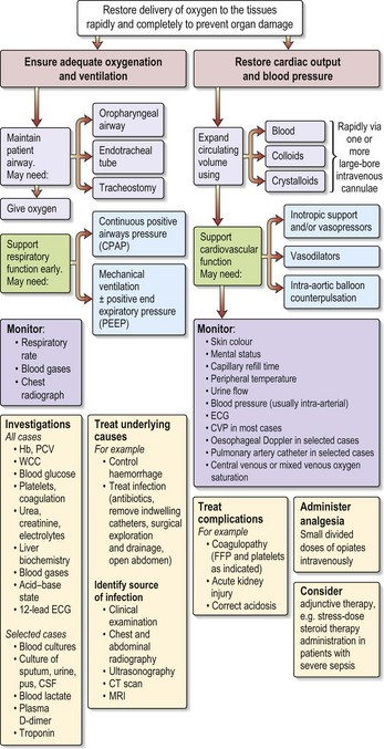
Other investigations (see Fig 12.2)
• Arterial blood gas analysis: in hypovolaemic shock (as in all shock states) there is a metabolic acidosis with a high hydrogen ion concentration and low bicarbonate concentration. In cases with respiratory complications the pO2 and pCO2 values will help indicate the need for ventilatory support.
• Lactate levels: blood lactate levels rise approximately in proportion to the severity of the shock.
• X-rays: these are usually of little value in the acute stages of shock management. A CXR will exclude any treatable pathology, such as a pneumothorax or haemothorax often complicating insertion of monitoring lines. A CT scan may be helpful in, for example, a case of trauma.
How would you treat?
Both patients should be admitted to high-dependency nursing units in the first instance as their MEWS scores were > 5.
Case 1
In cardiogenic hypotension key issues are pain relief, arrhythmia management and treatment of pulmonary oedema. Pain relief by incremental doses of intravenous opiates will aid reduction in myocardial oxygen consumption. Correcting electrolyte disturbances, hypoxia and controlling angina pain might assist in arrhythmia management. Temporary transvenous pacing might be required for significant bradycardia. Continuous infusion of vasodilators (e.g. glyceryl trinitrate 10–200 µg/min) plus diuretics (e.g. furosemide 40 mg) is needed for pulmonary oedema. Acute revascularisation (thrombolysis, angioplasty) might also be indicated. In the context of persistent hypotension, infusion of inotropic agents such as dobutamine (e.g. 2.5–10 µg/kg/min) might be necessary whilst correctable abnormalities are sought (e.g. acute mitral regurgitation following papillary muscle rupture or the development of an ischaemic ventricular septal defect). Percutaneous insertion of an intra-aortic balloon counterpulsation pump may be necessary for refractory cardiogenic hypotension following transfer to a specialist centre as a prelude to surgical intervention.
Progress.
This man initially improved on ionotropes, diuretics and vasodilatory therapy. However, he remained hypotensive, BP < 90 with poor output. He was transferred to the Cardiac Centre for insertion of an intra-aortic balloon pump. He did not respond and died 3 days later (mortality is 79% for cardiogenic shock).
Information
Patients at risk of further deterioration will have one or more of the following vital signs, which would indicate transfer to an intensive therapy unit (ITU):
• Systolic blood pressure > 200 mmHg
• Systolic blood pressure < 80 mmHg
• Respiratory rate > 30 breaths per minute
• Respiratory rate < 8 breaths per minute
• Urine output less than 0.5 mL/kg/h for 2 consecutive hours.

Stay updated, free articles. Join our Telegram channel

Full access? Get Clinical Tree

