1 Introduction to pharmacology
After reading this chapter, you will:
Molecular basis of pharmacology
What is pharmacology?
Pharmacology is the study of the actions, mechanisms, uses and adverse effects of drugs.
A drug is any natural or synthetic substance that alters the physiological state of a living organism. Drugs can be divided into two groups:
Although drugs are intended to have a selective action, this is rarely achieved.
There is always a risk of adverse effects associated with the use of any drug and the prescriber should weigh up the effects when choosing drugs.
Drug names and classification
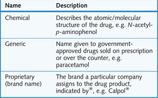
A single drug can have a variety of names (Fig. 1.1) and belong to many classes. Drugs are classified according to their:
When a drug company’s patent expires, the marketing of the drug is open to other manufacturers. Although the generic name is retained the brand names can be changed.
How do drugs work?
Most drugs produce their effects by targeting specific cellular macromolecules. The majority act on receptors but they can also inhibit enzymes and transport systems. Some drugs directly target pathogens. For example, β-lactam antibiotics are bactericidal, acting by interfering with bacterial cell wall synthesis.
Certain drugs do not have conventional targets. For example, succimer is a chelating drug that is used to treat heavy metal poisoning. It binds to metals, rendering them inactive and more readily excretable. Such drugs work by means of their physicochemical properties and are said to have a non-specific mechanism of action. For this reason these drugs must be given in much higher doses (mg–g) than the more specific drugs.
Transport systems
Ion channels
Ion channels are proteins that form pores across the cell membrane and allow selective transfer of ions (charged species) in and out of the cell. Opening or closing of these channels is known as ‘gating’; this occurs as a result of the ion channel undergoing a change in shape. Gating is controlled either by transmitter substances or by the membrane potential (voltage-operated channels).
Some drugs modulate ion channel function directly by blocking the pore (e.g. the blocking action of local anaesthetics on sodium channels); others bind to a part of the ion channel protein to modify its action (e.g. anxiolytics acting on the GABA channel). Other drugs interact with ion channels indirectly via a G-protein and other intermediates.
Carrier molecules
Transfer of ions and molecules against their concentration gradients is facilitated by carrier molecules located in the cell membrane. There are two types of carrier molecule:
Enzymes
Enzymes are protein catalysts that increase the rate of specific chemical reactions without undergoing any net change themselves during the reaction. All enzymes are potential targets for drugs. Drugs either act as a false substrate for the enzyme or inhibit the enzyme’s activity directly by binding to other sites on the enzyme.
Certain drugs may require enzymatic modification. This degradation converts a drug from its inactive form (prodrug) to its active form.
Receptors
Receptors are the means through which endogenous ligands produce their effects. A receptor is a specific protein molecule that is usually located in the cell membrane, although intracellular receptors and intranuclear receptors also exist.
A ligand that binds and activates a receptor is an agonist. However, a ligand that binds to a receptor but does not activate the receptor, also prevents an agonist from doing so. Such a ligand is called an antagonist.
The following are naturally occurring ligands:
Each cell expresses only certain receptors, depending on the function of the cell. Receptor number and responsiveness to messengers can be modulated.
In many cases there is more than one receptor for each messenger, so that the messenger often has different pharmacological specificity and different functions according to where it binds (e.g. adrenaline is able to produce different effects in different tissues).
Using conventional molecular biology techniques it is now possible to clone receptors and express them in cultured cells, thus allowing their properties to be studied. In particular, amino acid mutations can be reproduced so that the relation between protein structure and function can be evaluated.
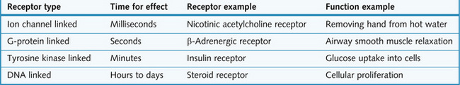
There are four main types of receptor (Fig. 1.2).
1 Receptors directly linked to ion channels
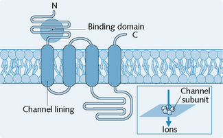
Receptors that are directly linked to ion channels (Fig. 1.3) are mainly involved in fast synaptic neurotransmission. A classic example of a receptor linked directly to an ion channel is the nicotinic acetylcholine receptor (nicAChR).

Fig. 1.3 General structure of the subunits of receptors directly linked to ion channels. (C, C-terminal; N, N-terminal.)
(Redrawn from Page et al. 2006.)
The nicAChRs possess several characteristics:
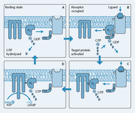
2 G-protein linked receptors
G-protein linked receptors (Fig. 1.4) are involved in relatively fast transduction. G-protein linked receptors are the predomínant receptor type in the body; muscarinic, ACh, adrenergic, dopamine, serotonin and opiate receptors are all examples of G-protein linked receptors.

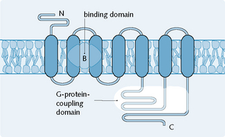
Fig. 1.4 General structure of the subunits of receptors linked to G-proteins. (C, C-terminal; N, N-terminal.)
(Redrawn from Page et al. 2006.)
Molecular structure of the receptor
Most of the G-protein linked receptors consist of a single polypeptide chain of 400–500 residues and have seven transmembrane-spanning α helices. The third intracellular loop of the receptor is larger than the other loops and interacts with the G-protein.
The ligand-binding domain is buried within the membrane on one or more of the α helical segments. In contrast to the ion channel coupled receptors, the ligand binds to the extracellular N-terminal region – an area easily accessible to small hydrophobic molecules.
G-proteins
Figure 1.5 illustrates the mechanism of G-protein linked receptors:

Fig. 1.5 Mechanism of action of G-protein linked receptors. (α, β, γ, subunits of G-protein; ATP, adenosine triphosphate; cAMP, cyclic adenosine monophosphate; G, guanosine; GDP, GTP, guanosine di- and triphosphate; p, phosphate.)
(Redrawn from Page et al. 2006.) Integrated Pharmacology, 3rd edition, Mosby.
This whole process results in an amplification effect because the binding of an agonist to the receptor can cause the activation of numerous G-proteins which in turn can each, via their association with the effector, produce many molecules of product.
Many types of G-protein exist. This is probably attributable to the variability of the α subunit. Gs and Gi/Go cause stimulation and inhibition, respectively, of the target enzyme adenylyl cyclase. This explains why muscarinic ACh receptors (Gi/Go linked) and β-adrenoreceptors (Gs linked) located in the heart produce opposite effects. The bacterial toxins cholera and pertussis can be used in order to determine which G-protein is involved in a particular situation. Each has enzymic action on a conjugation reaction with the α subunit, such that:
Targets for G-proteins
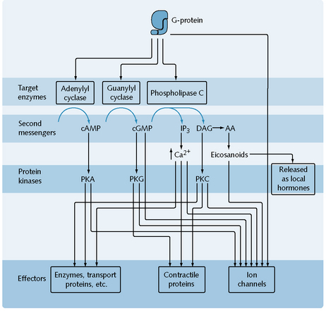
G-proteins interact with either ion channels or secondary messengers. G-proteins may activate ion channels directly, e.g. muscarinic receptors in the heart are linked to potassium channels which open directly on interaction with the G-protein, causing a slowing down of the heart rate. Secondary messengers are a family of mediating chemicals that transduces the receptor activation in to a cellular response. These mediators can be targeted and three main secondary messenger systems exist as targets of G-proteins (Fig. 1.6).

Fig. 1.6 Second-messenger targets of G-proteins and their effects. (AA, arachidonic acid ; cAMP, cyclic adenosine monophosphate; cGMP, cyclic guanosine monophosphate; DAG, diacylglycerol; IP3, inositol (1,4,5) triphosphate; PK, protein kinase.)
Adenylyl cyclase/cAMP system
Adenylyl cyclase catalyses the conversion of ATP to cyclic cAMP within cells. The cAMP produced in turn causes activation of certain protein kinases, enzymes that phosphorylate serine and threonine residues in various proteins, thereby producing either activation or inactivation of these proteins. A stimulatory example of this system can be observed in the activation of β1-adrenergic receptors found in cardiac muscle. The activation of β1-adrenergic receptors results in the activation of cAMP-dependent protein kinase A, which phosphorylates and opens voltage-operated calcium channels. This increases calcium levels in the cells and results in an increased rate and force of contraction. An inhibitory example of this system can be observed in activation of opioid receptors. The receptor linked to the ‘Gi’ protein inhibits adenylyl cyclase and reduces cAMP production.
Phospholipase C/inositol phosphate system
Activation of M1, M3, 5-HT2, peptide and α1-adrenoreceptors, via Gq, cause activation of phospholipase C, a membrane-bound enzyme, which increases the rate of degradation of phosphatidylinositol (4,5) bisphosphate into diacylglycerol (DAG) and inositol (1,4,5) triphosphate (IP3). DAG and IP3 act as second messengers. IP3 binds to the membrane of the endoplasmic reticulum, opening calcium channels and increasing the concentration of calcium within the cell. Increased calcium levels may result in smooth muscle contraction, increased secretion from exocrine glands, increased hormone or transmitter release, or increased force and rate of contraction of the heart. DAG, which remains associated with the membrane owing to its hydrophobicity, causes protein kinase C to move from the cytosol to the membrane where DAG can regulate the activity of the latter. There are at least six types of protein kinase C, with over 50 targets including:
Guanylyl cyclase system
Guanylyl cyclase catalyses the conversion of GTP to cGMP. This cGMP goes on to cause activation of protein kinase G which in turn phosphorylates contractile proteins and ion channels. Transmembrane guanylyl cyclase activity is exhibited by the atrial natriuretic peptide receptor upon the binding of atrial natriuretic peptide. Cytoplasmic guanylyl cyclase activity is exhibited when bradykinin activates receptors on the membrane of endothelial cells to generate nitric oxide, which then acts as a second messenger to activate guanylyl cyclase within the cell.
3 Tyrosine kinase linked receptors
Tyrosine kinase linked receptors are involved in the regulation of growth and differentiation, and responses to metabolic signals. The response time of enzyme-initiated transduction is slow (minutes). Examples include the receptors for insulin, platelet-derived growth factor and epidermal growth factor.
Activation of tyrosine kinase receptors results in autophosphorylation of tyrosine residues leading to the activation of pathways involving protein kinases.
4 DNA linked receptors
DNA linked receptors are located intracellularly and so agonists must pass through the cell membrane in order to reach the receptor. The agonist binds to the receptor and this receptor–agonist complex is transported to the nucleus, aided by chaperone proteins. Once in the nucleus the complex can bind to specific DNA sequences and so alter the expression of specific genes. As a result, transcription of this specific gene to mRNA is increased or decreased and thus the amount of mRNA available for translation into a protein increases or decreases. The process is much slower than for other receptor–ligand interactions, and the effects usually last longer. Examples of molecules with DNA-linked receptors are corticosteroids, thyroid hormone, retinoic acid and vitamin D.
Drug–receptor interactions
Most drugs produce their effects by acting through specific protein molecules called receptors.
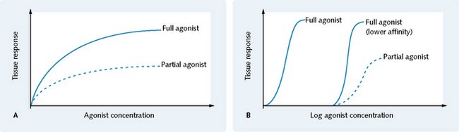
Receptors respond to endogenous chemicals in the body that are either synaptic transmitter substances (e.g. ACh, noradrenaline) or hormones (endocrine, e.g. insulin; or local mediators, e.g. histamine). These chemicals or drugs are classed as:
Electrostatic forces initially attract a drug to a receptor. If the shape of the drug corresponds to that of the binding site of the receptor, then it will be held there temporarily by weak bonds or, in the case of irreversible antagonists, permanently by stronger covalent bonds. It is the number of bonds and goodness of fit between drug and receptor that determines the affinity of the drug for that receptor, such that the greater the number of bonds and the better the goodness of fit, the higher the affinity will be.
The affinity is defined by the dissociation constant, which is given the symbol Kd. The lower the Kd, the higher the affinity. Kd values in the nanomolar range represent drugs (D) with a high affinity for their receptor (R):
The rate at which the forward reaction occurs depends on the drug concentration [D] and the receptor concentration [R]:
The rate at which the backward reaction occurs mainly depends on the interaction between the drug and the receptor [DR]:
Ka is the association constant and is used to quantify affinity. It can be defined as the concentration of drug that produces 50% of the maximum response at equilibrium, in the absence of receptor reserve:
Drugs with a high affinity stay bound to their receptor for a relatively long time and are said to have a slow off-rate. This means that at any time the probability that any given receptor will be occupied by the drug is high.
The ability of a drug to combine with one type of receptor is termed specificity. Although no drug is truly specific, most exhibit relatively selective action on one type of receptor.
Agonists
Agonist (A) binds to the receptor (R) and the chemical energy released on binding induces a conformational change that sets off a chain of biochemical events within the cell, leading to a response (AR*). The equation for this is:
where: (1) affinity; (2) efficacy.
Partial agonists cannot bring about the same maximum response as full agonists, even if their affinity for the receptor is the same (Fig. 1.7).

Fig. 1.7 Comparison for a partial agonist and a full agonist showing (A) the dose–response curve and (B) the log dose–response curve.
(From Neal MJ 2009 Medical Pharmacology at a Glance, 6th edition. Wiley-Blackwell, with permission.)

Stay updated, free articles. Join our Telegram channel

Full access? Get Clinical Tree

