Introduction to Genetic Diseases

Chapter 9 Introduction to Genetic Diseases


In theory, one half of the genes in diploid somatic cells should be identical to genes in the father, the other half should be identical to genes in the mother, and the genes should be identical in all cells of the body. In reality, they are not—not quite. Maintaining a bloated genome of three billion base pairs is such a formidable task that replication errors and other molecular accidents are unavoidable. These errors are called mutations, and they are a major cause of disease and disability.


Somatic mutations can produce cells with reduced viability or impaired function. They accumulate with age and contribute to normal aging. The most dangerous somatic mutations are those that cause the cell to grow out of control. Mutations of this type are the principal cause of cancer, which is responsible for 20% of all deaths in the modern world. This implies that all mutagenic agents are carcinogenic.


Germline mutations arise in the gametes or their diploid ancestors in the gonads. They are transmitted to the offspring and can cause genetic diseases.


This chapter introduces the various types of mutation, their importance for disease, and the DNA repair systems that the body uses to protect itself against mutations. The chapter also presents the hemoglobinopathies as examples of genetic diseases with well-understood pathogenesis.


Four types of genetic disease


Four types of genetic disease are commonly distinguished.


1. Aneuploidy is an aberration in chromosome number, caused by faulty segregation of chromosomes during mitosis or meiosis. About 1 in 400 infants is born aneuploid. In trisomy 21 (Down syndrome), for example (Fig. 9.1), one of the smallest autosomes (non-sex chromosomes) is present in three rather than the usual two copies. Presumably the signs of Down syndrome are caused by overproduction of the proteins encoded by the 225 genes on chromosome 21. Most cases of aneuploidy originate in female meiosis I, and the risk rises with advanced maternal age.


CLINICAL EXAMPLE 9.1: Paternal Age and Schizophrenia


About 1% of all people are diagnosed with schizophrenia at one or another time in their lives. This “multifactorial” disease manifests with delusions, hallucinations, and other thought disorders. The prevalence of schizophrenia is similar in all human populations, and its heritability is 60% to 80%. How can “schizophrenia genes” (actually, genetic variants that increase schizophrenia risk) be maintained at such high frequencies that they turn 1% of the population insane in each generation?


In most places, schizophrenics have fewer children than the average in the population. Unaffected relatives of schizophrenics, who are expected to carry some of the predisposing genes without expressing the disease, have no more children than everyone else. Therefore the offending genes should be eliminated by natural selection, but they are not.


The reason is shown by the observation that the fathers of schizophrenics are, on average, a few years older than the fathers of unaffected people. Maternal age has no independent effect. We know that new mutations leading to dominant diseases are more common in the children of older fathers because replication errors accumulate in the paternal germ line with advancing age. The spermatogonia of a 15-year-old boy have gone through an estimated 35 mitoses, but those of a 50-year-old man have gone through 800.


Molecular genetic studies are beginning to show that genetic liability to schizophrenia is caused by individually rare mutations in a fairly large number of genes. The effects of new mutations add to those of inherited mutations, raising the disease risk for the children of old fathers. The lesson for women is this: Take a young man rather than an old man as the father of your children!


Small mutations lead to abnormal proteins


Base substitutions in the coding sequences of genes are responsible for about 60% of disease-causing mutations, and small insertions and deletions cause another 20% to 25%. Less than 1% of single-gene disorders are caused by a mutation in a regulatory site.


A point mutation is a change in a single base pair of the DNA. It is called transition if a purine is replaced by another purine or a pyrimidine by another pyrimidine, and it is called transversion if a purine is replaced by a pyrimidine or a pyrimidine by a purine.


In the coding sequence of a gene, the most common consequence of a point mutation is a single amino acid substitution in the polypeptide. For example:


image


image


This is called a missense mutation. Some missense mutations leave the biological functions of the protein intact, but others destroy them partially or completely. Synonymous mutations, also called silent mutations, are point mutations that do not change an amino acid because they create a new codon that still codes for the same amino acid. For example:


image


image


A nonsense mutation generates a premature stop codon. It causes the premature termination of translation, usually with the complete loss of function in the truncated protein. For example:


image


image


A frameshift mutation is caused by a small insertion or deletion. Although the amino terminal portion of the encoded protein is normal, the amino acid sequence is garbled beyond the site of the mutation because the messenger RNA (mRNA) is translated in the wrong reading frame. The protein product is most likely nonfunctional. For example:


image


image


However, the insertion or deletion of three base pairs, or any multiple of three, does not result in a frameshift.


Splice-site mutations change an intron-exon junction or the branch site within the intron. They cause the synthesis of an abnormally spliced protein.


Promoter mutations, as well as mutations in other regulatory sites, leave the structure of the polypeptide intact but change its rate of synthesis.


The basal mutation rate is caused mainly by replication errors


The basal mutation rate, which is observed in the absence of environmental mutagens, is caused mainly by errors during DNA replication. These replication errors have an important consequence. Because the number of mitotic divisions before formation of the gametes is far greater for spermatogonia than oogonia, most base substitutions and small insertions and deletions originate in the paternal rather than the maternal germ line (see Clinical Example 9.1).


Spontaneous tautomeric shifts in the bases contribute to replication errors. For example, thymine normally is present in the keto form and pairs with adenine. Very rarely, however, it shifts spontaneously to the enol form, which pairs with guanine. If a thymine in the template strand happens to be in the rare enol form at the moment of DNA replication, G instead of A is incorporated in the new strand.


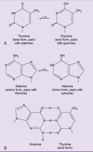
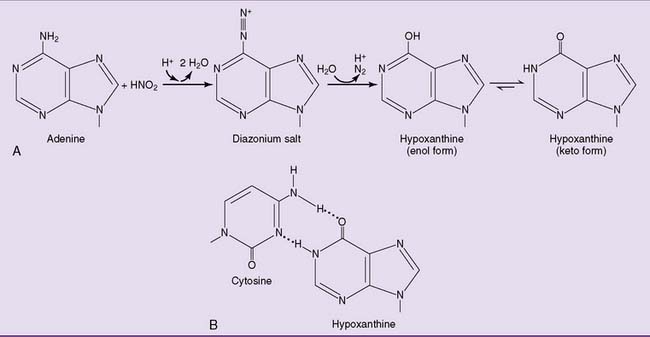
Similarly, adenine has a rare imino form that pairs with cytosine rather than thymine (Fig. 9.3). Fortunately, these bases spend very little time in their less stable forms; thus, mutations caused by tautomeric shifts are rare.


Mutations are also caused by short-lived, highly reactive free radicals that are formed during oxidative reactions in the cell, including superoxide and hydroxyl radicals. Free radicals cause strand breaks and oxidation of bases in DNA. They appear to be most important for mutagenesis in mitochondrial DNA.


Mutations can be induced by radiation and chemicals


Radiation is an avoidable cause of mutations. Ionizing radiation, including x-rays and radioactive radiation, is sufficiently energy rich to displace electrons from their orbitals. It damages DNA both directly, and indirectly through the formation of highly reactive hydroxyl radicals from water molecules. DNA double-strand breaks are the most important type of damage caused by ionizing radiation. Ionizing radiation penetrates the whole body and therefore can cause both somatic and germline mutations.


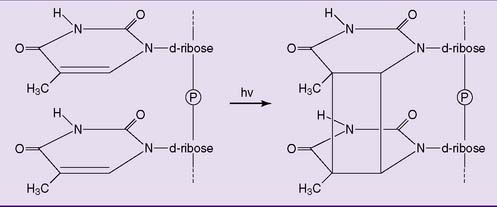
Ultraviolet radiation is a mutagenic component of sunlight. It cannot penetrate beyond the outer layers of the skin and therefore is unable to cause germline mutations. It only causes sunburn and skin cancer, mainly through the formation of pyrimidine dimers (Fig. 9.4).


Many chemicals can act as mutagens.
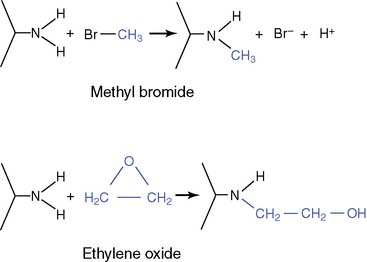


Mutagens are most mutagenic during the S phase of the cell cycle because mutagenesis during S phase leaves no time for repair of damaged DNA. This is the rationale for radiation treatment of cancer. Cancer cells divide more frequently than normal cells and therefore more likely are in S phase when the radiation is applied.


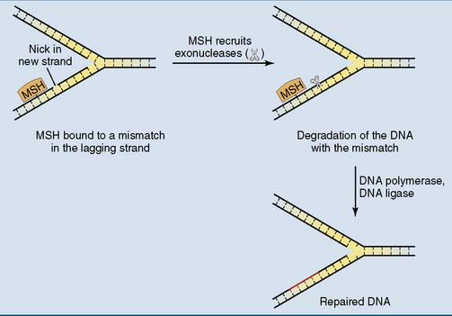
Mismatch repair corrects replication errors


DNA repair is required as part of life’s perennial struggle against the second law of thermodynamics (that entropy tends to rise over time). To maintain the genome, the repair enzymes have to proceed like a plumber who repairs a damaged pipe: Locate the damage, remove the damaged part, and replace it with a good part. Because of the great diversity of lesions that are generated in DNA every day, multiple repair systems with overlapping specificities are required.


Base mismatches that arise as replication errors pose a special problem for repair because the repair enzymes must distinguish between the intact old strand and the mutated new strand. Therefore postreplication mismatch repair requires at least two components: one to recognize the mismatch and the other to distinguish between the strands.


The new strand is distinguished from the old by the presence of frequent nicks. In the lagging strand, the nicks are present from the beginning until the Okazaki fragments are sealed by DNA ligase. However, even the leading strand is known to have occasional nicks.


The bound repair proteins recruit exonucleases to the nick, which then remove the DNA of the new strand between the nick and the mismatch, including the mismatch itself. This sets the stage for DNA polymerase δ and DNA ligase to fill the gap and connect the loose ends (Fig. 9.7). This system is most important for rapidly dividing cells (Clinical Example 9.2).


Jun 18, 2016 | Posted by in BIOCHEMISTRY | Comments Off on Introduction to Genetic Diseases

Full access? Get Clinical Tree

Get Clinical Tree app for offline access