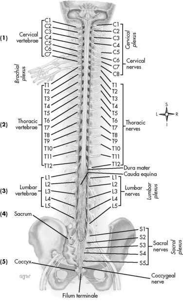
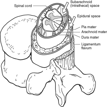
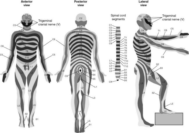
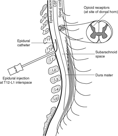
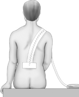
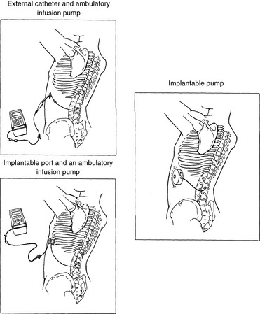
Intraspinal Analgesia (Epidural and Intrathecal)

Chapter 15


Intraspinal Analgesia (Epidural and Intrathecal)


Chapter outline


THE term intraspinal refers to the spaces or potential spaces surrounding the spinal cord or the nerve roots that constitute the cauda equina. Most often, the term is used when referring to the epidural and intrathecal spaces, each of which offers a route of administration for medications. The word neuraxial also is used to describe the group of spaces into which analgesic drugs can be administered. The word spinal is used interchangeably with the word intrathecal when referring to route of administration. It may also be used when referring generally to all of the routes of administration near or within the spinal meninges (Swarm, Karanikolas, Cousins, 2004). Intrathecal is often used synonymously with subarachnoid, but anatomically the intrathecal space includes the subdural space (Swarm, Karanikolas, Cousins, 2004) (see the following paragraphs on spinal anatomy). Table 15-1 shows some of the persistent misconceptions related to epidural analgesia. Box 15-1 presents patient selection guidelines and considerations for intraspinal analgesia.


Guidelines


Box 15-1   Use of Intraspinal Analgesia


Patient selection


• Absence of contraindications to intraspinal needle or catheter placement (e.g., coagulopathies, abnormal clotting studies, immunocompromise, sepsis or signs of systemic infection [elevated white cells, pyrexia], infection in region of puncture site, history of multiple abscesses, patient refusal).


• For patients with persistent cancer or noncancer pain, pain is uncontrolled and/or adverse effects are unmanageable and intolerable with systemic analgesics.


• For patients with acute pain (e.g., surgery, trauma), the systemic routes have been considered and are not an option because they would produce unmanageable and intolerable adverse effects at the anticipated doses required for adequate analgesia.


• Patient has a painful condition or surgical procedure for which reduced morbidity and mortality is important but impractical or unattainable with other routes of administration. Such conditions include major thoracic, abdominal, and orthopedic surgery; intractable MI.


• Intraspinal preemptive analgesia could prevent or reduce the severity of a persistent postsurgical pain syndrome (e.g., phantom limb pain, postthoracotomy pain).


• Intraspinal route will be used to deliver anesthesia for surgery, and a single bolus dose before removal of needle or catheter will produce acceptable postoperative analgesia (e.g., preservative-free epidural morphine or extended-release epidural morphine for cesarean section or hip replacement).


• Patient has a pain syndrome that may be responsive to a specific intraspinal therapy such as local anesthetics, clonidine, or steroids (e.g., neuropathic pain unresponsive to systemic and topical adjuvant analgesics).


• In the patient with cancer or persistent noncancer pain who will receive long-term intraspinal opioid therapy, a reduction in pain in response to a trial dose of an intraspinally administered opioid has been experienced.


From Pasero, C., & McCaffery, M. Pain assessment and pharmacologic management, p. 406, St. Louis, Mosby. Data from Brown, D. L. (2005). Spinal, epidural, and caudal anesthesia. In R. D. Miller (Ed.), Miller’s anesthesia, vol 2, ed 6, Philadelphia, Elsevier; Cashman, J. (2008). Routes of administration. In P. E. Macintyre, S. M. Walker, & D. J. Rowbotham (Eds.), Clinical pain management. Acute pain, ed 2, London, Hodder Arnold; Cousins, M. J., & Veering, B. T. (1998). Epidural neural blockade. In M. J. Cousins, & P. O. Bridenbaugh (Eds.), Neural blockade in clinical anesthesia and management of pain, Philadelphia, Lippincott-Raven; Swarm, R. A., Karanikolas, M., & Cousins, M. J. (2004). Anesthetic techniques for pain control. In D. Doyle, G. Hanks, N. I. Cherny, et al. (Eds.), Oxford textbook of palliative medicine, ed 3, New York, Oxford Press; Vascello, L., & McQuillan, R. J. (2006). Opioid analgesics and routes of administration. In O. A. de Leon-Casasola (Ed.), Cancer pain. Pharmacological, interventional and palliative care approaches, Philadelphia, Saunders; Wedel, D. J., & Horlocker, T. T. (2006). Regional anesthesia in the febrile or infected patient. Reg Anesth Pain Med, 31(4), 324-333. Pasero C, McCaffery M. May be duplicated for use in clinical practice.


Table 15-1


Misconceptions: Epidural Analgesia


Misconception Correction
Compared with opioid administration via IM injection and IV PCA, the incidence of respiratory depression is higher when opioids are administered by the epidural route. The incidence of respiratory depression associated with the various pain control methods is not firmly established because of a lack of consensus on definitions and well-controlled research, but the incidence of respiratory depression with epidural analgesia is less than that of IM opioid injections and probably more consistent with that of IV PCA. A systematic review of the literature concluded that the mean reported incidence of opioid-induced respiratory depression varied between 0.8% and 37.0% for IM injection; 1.2% and 11.5% for IV PCA; and 1.1% and 15.0% for epidural analgesia (Cashman, Dolin, 2004). A study of the use of PCEA morphine with basal rate or IV PCA morphine with basal rate in 2696 patients after major surgery reported a higher incidence of respiratory depression with IV PCA (1.2%) than epidural analgesia (0.04%) (Flisberg, Rudin, Linner, et al., 2003). Clinically significant opioid-induced respiratory depression can be avoided in opioid-naïve patients by slow titration, careful nurse monitoring of sedation levels and respiratory status, and decreases in opioid dose when increased sedation is detected (see Chapter 19).
Patients receiving epidural analgesia must be cared for in intensive care settings where their respiratory status can be mechanically monitored. Patients receiving epidural analgesia have been cared for safely outside of the intensive care setting for many years. Though mechanical monitoring is warranted in patients at high risk for respiratory complications (e.g., those with obstructive sleep apnea, chronic pulmonary disease), nurse assessment of sedation level and respiratory status is reliable and the most common method for monitoring most patients receiving epidural analgesia (see Chapter 19).
Epidural local anesthetics cause excessive and disabling sensory and motor blockade. Local anesthetics are administered in low (subanesthetic) doses (e.g., 0.05% to 0.125% bupivacaine; 0.1% to 0.2% ropivacaine) for epidural analgesia. Higher doses are required to produce significant motor and sensory blockade (0.5% to 0.75% bupivacaine; 0.75 to 1.0% ropivacaine). Patients receiving epidural analgesia are able to ambulate and perform all the routine recovery activities expected of them to the extent their medical or surgical condition allows. The occasional occurrence of minor temporary numbness of lower extremities is resolved easily by decreasing the dose or removing the local anesthetic from the epidural analgesic solution.
Thoracic epidural catheter placement is technically more difficult and causes more damage than lumbar catheter placement. The technique for placing a thoracic epidural catheter is quickly mastered by anesthesia providers. A review of 874 cases of high thoracic epidural analgesia provided over a 7-year period revealed no related neurologic complications (Royse, Soeding, Royse, 2007).

IM, Intramuscular; IV, intravenous; PCA, patient-controlled analgesia.


In spite of widespread use, misconceptions related to epidural analgesia persist. This table corrects some of these misconceptions.


From Pasero, C., & McCaffery, M. Pain assessment and pharmacologic management, p. 405, St. Louis, Mosby. Data from American Society of Anesthesiologists Task Force on Neuraxial Opioids. (2009). Practice guidelines for the prevention, detection, and management of respiratory depression associated with neuraxial opioid administration. Anesthesiology, 110(2), 218-230; Brown, D. L. (2005). Spinal, epidural, and caudal anesthesia. In R. D. Miller (Ed.), Miller’s anesthesia, vol 2, ed 6, Philadelphia, Elsevier; Cashman, J. N., & Dolin, S. J. (2004). Respiratory and haemodynamic effects of acute postoperative pain management: Evidence from published data. Br J Anaesth, 93(2), 212-223; Cousins M. J., & Veering, B. T. (1998). Epidural neural blockade. In M. J. Cousins, & P. O. Bridenbaugh (Eds.), Neural blockade in clinical anesthesia and management of pain, Philadelphia, Lippincott-Raven; Dabu-Bondoc, S., Franco, S. A., & Sinatra, R. S. (2009). Neuraxial analgesia with hydromorphone, morphine, and fentanyl: Dosing and safety guidelines. In R. S. Sinatra, O. A. de Leon-Casasola, B. Ginsberg, et al. (Eds.), Acute pain management, Cambridge, NY, Cambridge University Press; Flisberg, P., Rudin, A., Linner, R., et al. (2003). Pain relief and safety after major surgery. A prospective study of epidural and intravenous analgesia in 2696 patients. Acta Anaesth Scand, 47(4), 457-465; Grape, S., & Schug, S. A. (2008). Epidural and spinal analgesia. In P. E. Macintyre, S. M. Walker, & D. J. Rowbotham (Eds.), Clinical pain management. Acute pain, ed 2, London, Hodder Arnold; Maalouf, D. B., & Liu, S. S. (2009). Clinical application of epidural analgesia. In R. S. Sinatra, O. A. de Leon-Casasola, B. Ginsberg, et al. (Eds.), Acute pain management, Cambridge, NY, Cambridge University Press; McCartney, C. J. L., & Niazi, A. (2006). Use of opioid analgesics in the perioperative period. In G. Shorten, D. B. Carr, D. Harmon, et al., (Eds.), Postoperative pain management: An evidence-based guide to practice, Philadelphia, Saunders; Royse, C. F., Soeding, P. F., & Royse, A. G. (2007). High thoracic epidural analgesia for cardiac surgery: An audit of 874 cases. Anaesth Intensive Care, 35(3), 374-377; Vascello, L., & McQuillan, R. J. (2006). Opioid analgesics and routes of administration. In O.A. de Leon-Casasola (Ed.), Cancer pain. Pharmacological, interventional and palliative care approaches, Philadelphia, Saunders. Pasero C, McCaffery M. May be duplicated for use in clinical practice.


Spinal Anatomy


The human spinal column consists of 33 individual vertebra referred to by their location: (1) 7 cervical, (2) 12 thoracic, (3) 5 lumbar, (4) 5 caudal or sacral (fused into one bone, the sacrum), and (5) 4 coccygeal (fused into one bone, the coccyx) (Figure 15-1).Vertebrae consist of an anterior body, the laminae that protect the lateral spinal cord, and spinous processes that project outwardly and posteriorly from the laminae. The vertebrae become larger as they descend in the vertebral column. The bones of the laminae are bound together by a number of ligaments (e.g., the dense ligamentum flavum) (Figure 15-2).


The spinal cord is located within and protected by the bony vertebral column and connective tissue (meninges). It is a continuous structure extending from the foramen magnum to approximately the first or second lumbar (L1 to L2) vertebral interspace. Below the tip of the spinal cord, which is called the conus medullaris, are the nerve roots that exit the spine from below the L2 vertebra to the lower part of the sacrum. This tangle of roots is known as the cauda equina.


Moving from outside to inside the spine, the epidural space is first encountered. This is a potential space filled with vasculature, fat, and a network of nerve extensions. No fluid is in the epidural space; a true space is created when volume or air is injected into it (see Figure 15-2). The epidural space is outside of the dura, which is composed of the dura mater and the arachnoid membranes. The subarachnoid space (also called the intrathecal space in the caudal part of the spine) lies deep to the subarachnoid membrane, between this membrane and the spinal cord and cauda equina. The subarachnoid space is filled with clear, colorless cerebrospinal fluid (CSF) that continually circulates and bathes the spinal cord and nerve roots.


The fact that the epidural space is a potential space has clinical implications. Although injecting large amounts of air is not recommended, small amounts, such as tiny bubbles within the infusion tubing when therapy is initiated, are not considered dangerous. In addition, because the epidural catheter is in a space and not a blood vessel, a continuous epidural infusion may be stopped for hours and restarted without concern that the catheter has become occluded. However, crystallization of the saline within the epidural catheter can occur when catheters are unused for prolonged periods. In these cases, weekly or biweekly irrigation is recommended (DuPen, DuPen, 1998).


At each vertebral body level, nerve roots exit from the spinal cord bilaterally. A specific area of skin and subcutaneous tissue, known as a dermatome, is innervated by a single spinal nerve (Figure 15-3). The assessment of sensation in a dermatome is used to determine the integrity of the nerve root and subsequent pathway of innervation. Assessment of sensation in dermatomes is performed by anesthesia providers and others to determine the level of spinal anesthesia for surgical procedures and postoperative analgesia when epidural local anesthetics are used.


Delivery of Intraspinal Analgesics


Delivery of analgesics by the intraspinal routes can be accomplished by inserting a needle into the subarachnoid space (for intrathecal analgesia) or the epidural space and injecting the analgesic, or threading a catheter through the needle and taping it in place temporarily for bolus dosing or continuous administration (Figures 15-4 to 15-6). Temporary catheters are used primarily for short-term acute pain management and are usually removed after 2 to 4 days. Intrathecal catheters for acute pain management are used more often for providing anesthesia and/or a single analgesic bolus dose.


For severe persistent cancer and noncancer pain, a catheter can be inserted then tunneled subcutaneously for intrathecal or epidural intermittent bolusing or continuous infusion or for patient-controlled epidural analgesia (PCEA) by an external ambulatory pump. The tunneling is done to decrease the incidence of infection and accidental displacement (Figure 15-7). These temporary tunneled catheters can be used for weeks to months to deliver analgesics. Temporary externalized intrathecal catheters are used less often than temporary epidural catheters primarily because of concerns about infection, although some clinicians report that such concerns may be unfounded (Vascello, McQuillan, 2006).


Although temporary tunneled epidural catheters continue to be useful for the management of intractable pain in some patients near end of life, totally implanted intrathecal infusion systems are preferred for long-term treatment of persistent pain (Deer, Krames, Hassenbusch, et al., 2007; Rathmell, Lake, Ramundo, 2006) (see Figure 15-7). Implanted catheters are less likely to dislodge and are associated with a lower infection rate than percutaneous catheters (Rathmell, Lake, Ramundo, 2006; Swarm, Karanikolas, Cousins, 2004) (see more on long-term intraspinal therapy later in the chapter).


The level of nociceptive input (e.g., surgical site, site of injury, tumor location), the characteristics of the opioid being administered, and the goals of care (e.g., reduced stress response) are most important in determining the vertebral level at which the catheter is placed (Maalouf, Liu, 2009). For example, long-term catheters for treatment of cancer pain associated with spinal lesions can be placed in a location that avoids the tumor while providing necessary analgesia (DuPen, DuPen, 1998). Temporary epidural catheters for acute pain management usually are placed at the lumbar or thoracic vertebral level depending on surgical site (see the section on dermatomal spread and catheter placement later in the chapter). For example, the high thoracic level is preferred by several clinicians for coronary artery bypass surgery because placement at this level improves coronary perfusion, decreases heart rate and endogenous stress response, and reduces the risk for myocardial ischemia (Kessler, Neidhart, Bremerich, et al., 2002; Paiste, Bjerke, Williams, et al., 2001; Royse, Royse, Soeding, et al., 2000).


Percutaneous Intraspinal Catheterization


Intraspinal needle and catheter insertion is performed usually by an anesthesiologist or certified registered nurse anesthetist (CRNA) or other advanced practice nurse. Nurses often assist with the procedure by preparing supplies and monitoring and supporting the patient during the procedure. Informed consent is obtained before the procedure.


The technique for placing a temporary percutaneous epidural catheter varies among practitioners; however, the points made in the Patient Example can be generalized to epidural catheter placement in all patients and may be helpful in reinforcing the anesthesia provider’s explanation of the procedure to patients. The same principles apply to intrathecal needle and catheter placement.


Patient Example


Mr. Z. and his wife want to know everything about the epidural catheter placement procedure he is going to receive later today. His nurse reinforces the anesthesia provider’s explanation by offering the following information: “You’ll either be in a sitting position or lying on your side for the procedure. The doctor will be behind you facing your back. First he’ll wash a small area on your back with a sponge. This may feel cool. Then he’ll put drapes on your back to keep the area as clean as possible. You’ll need to roll your shoulders inward and push your back out slightly. The doctor will use a local anesthetic to numb the place where the catheter will go. Sometimes injecting the local anesthetic produces a burning, stinging sensation that lasts less than a minute. It usually takes the doctor about 2 or 3 minutes to insert the needle into the epidural space. This will feel like dull pressure against your back. Next the doctor inserts the catheter through the needle into the epidural space, which usually takes less than a minute. As he inserts the catheter, you may feel some spark-like sensations in your legs and feet. These will go away very quickly. The doctor will remove the epidural needle and tape the catheter in place up your back to your shoulder. After the catheter is taped in place you’ll be able to move and turn and lie on your back like you did before the procedure. The whole procedure usually takes less than 30 minutes. You may feel a very slight irritation for an hour or two after the procedure just at the site where the catheter goes into your back. If you feel any more discomfort in your back than that at any time while the catheter is in place, you’ll need to let us know. Can I answer any questions?”


During intraspinal needle placement, most anesthesia providers are able to recognize when the point of the needle penetrates the dense ligamentum flavum (see Figure 15-2). In addition, entry into the epidural space exerts a negative pressure, which is registered by a loss of resistance in the syringe attached to the needle. Some anesthesia providers use the “hanging-drop” method whereby a drop of fluid at the needle hub is “sucked in” as soon as the needle tip passes the ligamentum flavum (Neruda, 2008); however, this method carries the risk of a small plug in the needle tip creating low or no negative pressure and is discouraged by some practitioners (Cousins, Veering, 1998). Once the ligamentum flavum is penetrated, the needle is not advanced if the epidural space is the desired location. If advanced further, the needle will penetrate the dura and enter the subarachnoid space. When in the subarachnoid space, free-flowing CSF can be aspirated. If a blood vessel is entered during placement, blood often can be aspirated.


Even when neither CSF nor blood is aspirated, epidural needle placement is often confirmed by injecting a test dose of lidocaine with epinephrine (if there is no contraindication to epinephrine; e.g., this approach is controversial in pregnant patients because of the potential difficulty in interpreting whether any variability in the woman’s heart rate is in response to epinephrine or to uterine blood flow and contractions; see Birnbach, Browne, 2005). If the needle is in a blood vessel, the epinephrine will cause the patient’s heart rate and blood pressure (BP) to increase suddenly and significantly; if in the subarachnoid space, the lidocaine will produce sensory anesthesia within 3 to 5 minutes (Covino, Wildsmith, 1998). If the patient exhibits neither of these changes, the needle is thought to be in the epidural space and the catheter is threaded through the needle.


Anesthesia providers turn the bevel of the intraspinal needle upward to facilitate threading the catheter 4 to 6 centimeters in a cephalad (toward the head) direction. Although rarely necessary for routine temporary intraspinal catheter placement, the only way to confirm conclusively the exact location of an intraspinal catheter is radiographically using contrast dye. When percutaneous catheters are to be used in the home setting, some clinicians recommend an epiduragram to confirm catheter position before patient discharge (DuPen, DuPen, 1998). (The reader is referred to a detailed explanation of epidural and intrathecal catheter placement techniques in the following two references: Brown, D. L. (2005). Spinal, epidural, and caudal anesthesia. In R. D. Miller (Ed.), Miller’s anesthesia, vol 2, ed 6, pp. 1653-1683, Philadelphia, Elsevier; and Cousins, M. J., & Veering, B. T. (1998). Epidural neural blockade. In M. J. Cousins, & P. O. Bridenbaugh (Eds.), Neural blockade in clinical anesthesia and management of pain, pp. 243-321, Philadelphia, Lippincott-Raven.


Intraspinal Analgesia for Persistent Cancer and Noncancer Pain


A systematic review of the literature in 2000 by a panel of experts revealed widespread acceptance of long-term intraspinal analgesia therapy despite a lack of scientific evidence to support it (Bennett, Serafini, Burchiel, et al., 2000). The need for more well-controlled research of this therapy continues today; most studies are retrospective and underpowered (Simpson, Jones, 2008). Another systematic review found reports of improvements in pain and function, but also remarked on methodologic problems with the studies in the review (Turner, Sears, Loeser, 2007). Another systematic review identified just 8 evaluable studies (177 patients) on long-term intraspinal analgesia, and all of the studies were described as low quality (Noble, Tregear, Treadwell, et al., 2008).


An early consensus guideline on long-term intrathecal analgesic drug delivery recommended morphine as the mainstay analgesic for long-term intrathecal pain management based on its long history of use, the panel’s extensive clinical experience with the drug, and responses to an online survey of physicians providing long-term intrathecal analgesia (Bennett, Burchiel, Buschser, et al., 2000). The survey revealed a usual starting morphine dose of 0.2 mg/day to 20 mg/day and an average maximum long-term infusion dose of 21.1 mg/day. Updated reviews of the literature and development of algorithms and dosing guidelines for the therapy were published in 2004 (Hassenbusch, Portenoy, Cousins, et al., 2004) and again in 2007 (Deer, Krames, Hassenbusch, et al., 2007). The 2007 recommendations list morphine, hydromorphone, and ziconotide as first-line options. Second-line choices included fentanyl alone, and combinations of morphine/hydromorphone plus ziconotide, or morphine/hydromorphone plus bupivacaine/clonidine. (See Section V for a detailed discussion of ziconotide and other agents administered for long-term intraspinal analgesia.) Table 15-2 provides concentrations and dosing recommendations from the most recent consensus guideline (Deer, Krames, Hassenbusch, et al., 2007).


The above-mentioned online survey found that drug and dose adjustments were common and that one-half of patients receiving long-term intrathecal pain management who began on a single drug eventually received polytherapy, indicating a common need to adjust therapy to improve pain control or reduce adverse effects (Hassenbusch, Portenoy, Cousins, et al., 2004). A review of the literature found that 6.3% of patients withdrew from clinical trials of long-term intrathecal therapy because of adverse effects, and 10.5% withdrew because of insufficient pain relief (Noble, Tregear, Treadwell, et al., 2008).


Some publications provide insight into the pros and cons of the therapy. A summary of responses to a questionnaire administered to 36 patients with persistent low back pain receiving long-term intrathecal opioid treatment (mean 4.38 years) revealed significant improvements in pain after spinal implantation and a nonsignificant trend toward enhanced quality of life (Raphael, Southall, Gnanadurai, et al., 2002). The majority (88%) thought the therapy was quite or very worthwhile, and only 1 patient (3%) responded that it was not worthwhile. A systematic review of six articles on effectiveness and four others on complications associated with programmable intrathecal opioid delivery systems for persistent noncancer pain concluded that the therapy produced improvements in pain and functioning, but the typical opioid-induced adverse effects and device complications, such as mechanical failure and catheter migration, were relatively common (Turner, Sears, Loeser, 2007). A Cochrane Collaboration Review concluded that controlled research is lacking on neuraxial analgesia for cancer treatment, and although the therapy is often effective for cancer pain that is unresponsive to systemic analgesia, intraspinal catheter complications frequently occur (Ballantyne, Carwood, 2005).


An excellent review of the literature identified complications associated with programmable intrathecal opioid delivery systems, which include infection (e.g., wound infection, meningitis), hardware problems (e.g., mechanical failure, catheter occlusion), and opioid-related adverse effects (Turner, Sears, Loeser, 2007). Life-threatening complications were rare. The most common adverse effects were nausea (33%), pruritus (26%), and urinary retention (24%). Only two studies evaluated effects on sexual function and reported a variety, including amenorrhea and erectile dysfunction. One case of opioid withdrawal syndrome from catheter disconnection was reported. See later in this chapter for a detailed discussion of complications during intraspinal therapy.


Cost is a major consideration when implanted pumps are used in patients who are terminally ill; according to an early cost-benefit study, an implanted infusion pump was more favorable when survival times exceeded 3 months (Bedder, Burchiel, Larson, 1991). Some consider the intrathecal route to be more efficient, capable of providing a better distribution of medication, and less expensive for cancer pain (Vascello, McQuillan, 2006). An important aspect of care is helping the patient and the patient’s family weigh all of the risks and benefits of long-term intraspinal analgesia therapy prior to initiation.


Stability and Compatibility of Agents for Analgesic Infusion Therapy


Research has established the stability and compatibility of admixtures of many of the commonly used agents for intraspinal and other infusion therapies. (For a discussion of microbiologic research on solutions, see the research list following this paragraph.) The reader is also referred to the Polyanalgesic Consensus Conference 2004 publication (Hassenbusch, Portenoy, Cousins, et al., 2004) for discussion of stability and compatibility of intraspinal analgesics. See the 2007 Polyanalgesic Consensus Conference recommendations for a detailed review of the research on the various drugs used for long-term intrathecal analgesia (Deer, Krames, Hassenbusch, et al., 2007).


• Baclofen (Alvarez, de Mazancourt, Chartier-Kastler, et al., 2004; Goodwin, Kim, Zuniga, 2001)


• Bupivacaine (Allen, Stiles, Wang, 1993; Classen, Wimbish, Kupiec, 2004; Hildebrand, Elsberry, Deer, 2001b; Nitescu, Hultman, Appelgren, et al., 1992; Rudich, Peng, Dunn, et al., 2004; Tu, Stiles, Allen, 1990; Wulf, Gleim, Mignat, 1994)


• Buprenorphine (Nitescu, Hultman, Appelgren, et al., 1992)


• Clonidine (Alvarez, de Mazancourt, Chartier-Kastler, et al., 2004; Classen, Wimbish, Kupiec, 2004; Goodwin, Kim, Zuniga, 2001; Hildebrand, Elsberry, Anderson, 2001b; Vranken, van Kan, van der Vegt, 2006; Wulf, Gleim, Mignat, 1994)


• Dexamethasone with ketamine (Watson, Lin, Morton, et al., 2005)


• Fentanyl (Allen, Stiles, Tu, 1990; Allen, Stiles, Wang, 1993; Chapalain-Pargade, Laville, Paci, et al., 2006; Nitescu, Hultman, Appelgren, et al., 1992; Tu, Stiles, Allen, 1990)


• Hydromorphone (Hildebrand, Elsberry, Anderson, 2001b; Rudich, Peng, Dunn, et al., 2004; Walker, Law, DeAngelis, 2001)


• Ketamine (Schmid, Koren, Klein, et al., 2002; Walker, Law, DeAngelis, 2001; Watson, Lin, Morton, et al., 2005)


• Meperidine (Nitescu, Hultman, Appelgren, et al., 1992; Vranken, van Kan, van der Vegt, 2006)


• Morphine (Classen, Wimbish, Kupiec, 2004; Hildebrand, Elsberry, Hassenbusch, 2003; Nitescu, Hultman, Appelgren, et al., 1992; Schmid, Koren, Klein, et al., 2002; Trissel, Pham, 2002; Trissel, Xu, Pham, 2002; Vermiere, Remon, 1999; Wulf, Gleim, Mignat, 1994)


• Ropivacaine (Sanchez del Aguila, Jones, Vohra, 2003)


• Sufentanil (Boitquin, Hecq, Evrard, et al., 2004; Chapalain-Pargade, Laville, Paci, et al., 2006)


• Tramadol with halodroperidol (Negro, Martin, Azuara, et al., 2005)


Methods for Administering Intraspinal Analgesia


The three methods for administering intraspinal analgesia are: (1) bolus (administered by the clinician), (2) continuous infusion or basal rate (administered by a pump), and (3) PCEA (administered by the patient usually using a pump).


Clinician-Administered Bolus Method


Clinicians can provide analgesia by administering a single intrathecal or epidural bolus injection, or the catheter can be left in place for intermittent bolus injections. The duration of the patient’s pain usually determines which bolus method is used.


For some surgical procedures, a single intraspinal morphine bolus provides sufficient pain control for several hours. For example, an epidural or intrathecal bolus of morphine often is administered to manage pain that does not warrant placement of a catheter, such as after cesarean section and some gynecologic, orthopedic, and urologic procedures (Dabu-Bondoc, Franco, Sinatra, 2009). A single epidural morphine dose is capable of providing analgesia for up to 24 hours to 48 hours depending on the formulation used (see the paragraphs that follow). After this period of time, pain usually can be controlled with oral or IV analgesics. Single bolusing is also used when continuous epidural infusions are contraindicated such as in some patients who require anticoagulant therapy (Dabu-Bondoc, Franco, Sinatra, 2009).


When moderate to severe pain is expected to be constant for more than 24 hours, the epidural catheter can be left in place to provide intermittent analgesic bolus doses; however, this method is rarely used today with advances in infusion devices by which to administer therapy that is required for more than 24 hours. As mentioned, when the intrathecal route is used for acute pain, analgesia is administered most often by single bolus; however, implanted subcutaneous ports can be accessed to deliver intermittent boluses for long-term intraspinal pain management. When an intrathecal catheter is implanted for long-term pain control, analgesia usually is provided by continuous infusion.


The major drawback of the intermittent epidural bolus method is that a steady analgesic level is difficult to maintain, especially when bolus doses are administered PRN. Relatively large doses of the opioid are given, and a “peak and trough” effect occurs. Patients experience adverse effects at the peak (highest analgesic concentration level) and pain at the trough (lowest analgesic concentration level). Rather than a PRN approach to epidural dosing, it may be preferable to consider smaller scheduled around-the-clock (ATC) doses. A dosing frequency of less than every 6 hours is not recommended (DuPen, DuPen, 1998) (see Box 15-2 for guidelines for administering intermittent boluses through a temporary epidural catheter).


Box 15-2   Intermittent Boluses via a Short-Term Epidural Catheter


• Administer only preservative-free solutions that are labeled safe for intraspinal use.


• Before injecting, verify with another RN that the preservative-free drug, dose, and volume are in accordance with anesthesia provider’s order.


• Before injecting, verify that the catheter to be injected is the epidural catheter. (Most catheters are color-coded to prevent errors, but this should always be checked prior to injection.)


• To prevent exerting too much pressure on injection, administer analgesic in at least a 10 mL syringe.


• The use of indwelling epidural filters depends on institutional policy (see text). A filter straw or needle should be used to draw solutions from glass ampules; if solutions are drawn unfiltered from glass ampules, they always should be injected through a 0.22 micron filter.


• Maintain sterility of the epidural system when removing catheter port cap, attaching syringes, and replacing port cap. (Some institutions require disinfecting connection before removing port cap. Use only nonneurotoxic agents to disinfect intraspinal catheter connections or ports [alcohol often is used to cleanse skin of secretions, but should not be used to disinfect catheter connections or ports].)


• Before injecting analgesic, gently aspirate catheter; allow time for fluid/air to travel up catheter1:


• Do not inject analgesic if patient reports pain on steady injection; notify anesthesia provider.


• Some resistance during injection is normal, but if strong resistance is met during injection, reposition the patient so that the spine is flexed. If resistance continues, stop and notify anesthesia provider.


• Do not flush epidural catheter after injecting analgesic unless specifically ordered to do so.


CSF, Cerebrospinal fluid.


When moderate to severe pain is expected to be constant for more than 24 hours, the epidural catheter is sometimes left in place to provide intermittent analgesic bolus doses. A majority of patients require epidural opioid bolus doses every 8 hours. Patients should be assessed systematically as frequency varies depending on analgesic requirement and opioid characteristics; some patients may require dosing more often, e.g., every 6 hours, while others may require boluses every 12 to 24 hours.


1Some anesthesia providers require the administration of a small test dose of local anesthetic (e.g., bupivacaine or ropivacaine) containing epinephrine to rule out intravascular or intrathecal migration prior to bolus administration; check with the state board of nursing before performing this function (also see Box 15-4 on p. 433).


From Pasero, C., & McCaffery, M. Pain assessment and pharmacologic management, p. 414, St. Louis, Mosby. Data from email communication and review on August 13, 2009, by Carol Mulvenon, MS, RN-BC, AOCN, ACHPN, Clinical Nurse Specialist, Pain Management-Palliative Care-Oncology, St. Joseph Medical Center, Kansas City, MO. Additional information from DuPen, S. L., & DuPen, A. R. (1998). Spinal analgesia. In M. A. Ashburn, & L. J. Rice (Eds.), The management of pain, New York, Churchill Livingstone; Grape, S., & Schug, S. A. (2008). Epidural and spinal analgesia. In P. E. Macintyre, S. M. Walker, & D. J. Rowbotham (Eds.), Clinical pain management. Acute pain, ed 2, London, Hodder Arnold; Maalouf, D. B., & Liu, S. S. (2009). Clinical application of epidural analgesia. In R. S. Sinatra, O. A. de Leon-Casasola, B. Ginsberg, et al. (Eds.), Acute pain management, Cambridge, NY, Cambridge University Press; Pasero, C., Eksterowicz, N., Primeau, M., et al. (2007). ASPMN position statement: Registered nurse management and monitoring of analgesia by catheter techniques. Pain Manage Nurs, 8(2), 48-54. Pasero C, McCaffery M. May be duplicated for use in clinical practice.


Continuous Infusion


The principle of providing continuous pain control with intraspinal analgesia can be accomplished by using an external (for acute pain and for persistent pain) or implanted (for persistent pain) infusion pump to deliver a continuous infusion (also called basal rate) of an analgesic solution. Supplemental bolus doses are prescribed for breakthrough pain and can be administered using the clinician-administered bolus mode available on most external infusion pumps or as outlined in Box 15-2. When implanted ports are used to deliver continuous infusion and/or intermittent boluses, meticulous aseptic precautions should be taken to protect the port from bacterial contamination (DuPen, DuPen, 1998; Holmfred, Vikerfors, Berggren, et al., 2006).


Continuous epidural analgesia has been shown to have more positive impact than IV PCA on some but not all patient outcomes following major surgery. One double-blind study randomized 60 patients undergoing radical prostatectomy to receive low thoracic (T10-T12) epidural ropivacaine (0.1%) plus fentanyl (2 mcg/mL) at 10 mL/h or IV PCA morphine (1 mg every 6 minutes) (Gupta, Fant, Axelsson, et al., 2006). Although there were no differences in hospital length of stay, those who received epidural analgesia had significantly better pain relief and expiratory muscle function than those who received IV PCA. Additionally, at 1 month, patients in the epidural group had better scores in emotional role, physical functioning, and general health. However, superior analgesia afforded by a continuous epidural infusion of bupivacaine and morphine in 60 older adults post–hip fracture surgery did not translate into improved rehabilitation in another randomized controlled study (Foss, Kristensen, Kristensen, et al., 2005). A prospective study showed similar results in 18 patients who received either epidural analgesia or IV PCA following mastectomy with immediate transverse rectus abdominis musculocutaneous (TRAM) flap breast reconstruction; continuous epidural analgesia produced better pain control and a 25-hour shorter hospital stay but no difference in time to first ambulation, first bowel sounds and flatus, tolerance of oral nutrition, and incidence of adverse effects (Correll, Viscusi, Grunwald, et al., 2001).


Patient-Controlled Epidural Analgesia (PCEA)


PCEA permits patients to treat their pain by self-administering doses of epidural analgesics to meet their individual analgesic requirements. A randomized controlled study compared fentanyl (4 mcg/mL) plus bupivacaine (0.125%) via PCEA (with basal rate) or continuous epidural infusion after colon resection and found that pain scores were similarly low but significantly fewer nurse/physician interventions for uncontrolled pain (e.g., epidural top-ups, systemic analgesia) were necessary, and patient satisfaction was significantly higher in those who received PCEA (Nightingale, Knight, Higgins, et al., 2007). Another randomized controlled study found that significantly less fentanyl-bupivacaine solution was consumed with PCEA (without basal rate) than with continuous epidural infusion following total knee arthroplasty (Silvasti, Pitkanen, 2001). Compared with nurse-administered PRN intermittent epidural bolus doses of meperidine (maximum of 50 mg/2 h), PCEA meperidine (25 mg PCEA dose with 10 minute lockout) resulted in better pain scores and a trend toward earlier return to activities of daily living and care for the newborn in women following cesarean section (Lim, Wilson, Katz, 2006). Although patient satisfaction was similar among the two groups, nurse satisfaction was higher with PCEA.


A retrospective review of the medical records of 245 patients who received PCEA (opioid plus local anesthetic) or IV PCA following lumbar spine surgery revealed that PCEA produced superior pain relief with less need for rescue analgesia (Cata, Noguera, Parke, et al., 2008). A randomized controlled study of older patients (N = 70) following major surgery observed better pain relief, higher patient satisfaction, and faster return of bowel function with PCEA (opioid plus local anesthetic) than with IV PCA (Mann, Pouzeratte, Boccara, et al., 2000). Although the incidence of postoperative delirium was similar among the two groups (24% to 26%), epidural analgesia was associated with improved mental status on the fourth and fifth day.


When PCEA is administered, a basal rate usually provides most of the patient’s analgesic requirement and the PCEA bolus doses are used to manage breakthrough pain. If a basal rate is not provided, it is especially important to remind patients to “stay on top of the pain,” to maintain a steady neuraxial analgesic level and self-administer bolus doses before pain is severe and out of control. Research has shown that this type of patient teaching is critical to successful therapy (Cywinski, Parker, Xu, et al., 2004). PCEA is safe and effective in older adults (Ishiyama, Iijima, Sugawara, et al., 2007; Mann, Pouzeratte, Boccara, et al., 2000), but the need for proper patient selection and frequent follow up to ensure appropriate PCEA use are emphasized (Silvasti, Pitkanen, 2001). See an example of a patient information brochure about PCEA on pp. 542-543 at the end of Section IV; see Chapter 12 for discussion of PCA principles and safeguards, such as patient-only use of PCA; and see Chapter 17 for discussion of PCA pump features and Table 17-2 on p. 469 for interventions for patients receiving PCEA.


Combined Spinal-Epidural Anesthesia/Analgesia (CSEA)


Although used less often than epidural analgesia, “combined spinal-epidural anesthesia/analgesia” (CSEA) is sometimes administered for labor and delivery and during and after cesarean section and other surgical procedures. CSEA involves placing an epidural needle (typically, 16- or 18-gauge Touhy) into the epidural space, and then passing a much smaller gauge and longer spinal needle (e.g., 29-gauge Quincke needle) through the epidural needle into the subarachnoid space. Subarachnoid placement is confirmed by aspiration of CSF. Opioid and/or local anesthetic is injected into the subarachnoid space, producing rapid and profound anesthesia/analgesia. The subarachnoid needle is removed, and an epidural catheter is inserted to administer supplemental doses as needed to prolong the block and provide ongoing analgesia (Cousins, Veering, 1998; Dabu-Bondoc, Franco, Sinatra, 2009).


The rationale for combining the routes is to minimize the shortcomings of both intrathecal and epidural analgesia while taking advantage of their benefits (Dabu-Bondoc, Franco, Sinatra, 2009; Teoh, Thomas, Tan, 2006). Intrathecal anesthesia has a rapid onset and produces dense neuraxial blockade, and epidural analgesia provides prolonged anesthesia and postoperative analgesia (Grape, Schug, 2008). For this reason, it has been suggested as an option for longer surgical procedures associated with significant postoperative pain, such as lower extremity surgery (Grape, Schug, 2008). CSEA improved intraoperative analgesia and reduced pain with cough better than an intermittent epidural bolus technique following major abdominal surgery in a prospective, randomized study (N = 160) (Stamenkovic, Geric, Slavkovic, et al., 2008) and produced faster motor recovery than single-shot spinal anesthesia following cesarean section in another prospective, randomized study (N = 62) (Lew, Yeo, Thomas, 2004). Because the CSEA technique uses the intrathecal route, lower doses of local anesthetic are possible for laboring patients, and less motor block is produced. The epidural route is used for low-dose supplemental analgesic boluses, and with appropriate assessment, patients have been able to safely and comfortably ambulate during labor while receiving CSEA (Brownridge, Cohen, Ward, 1998; Gautier, Debry, Fanard, et al., 1997).


Drug Bioavailability by the Intraspinal Routes


In contrast to drugs administered systemically, drugs administered intraspinally are more potent (i.e., small doses are effective) because distribution of the drug brings it close to the action site (opioid receptors in the dorsal horn of the spinal cord). This is particularly true when opioids are delivered by the intrathecal route where they are carried by the CSF to the dorsal horn. After epidural administration, drugs are distributed by three main pathways: (1) neural diffusion through the dura into the CSF then into the spinal cord directly to the receptors, (2) vascular uptake by the vessels in the epidural space into systemic circulation, and (3) uptake by the fat in the epidural space; a drug depot is created from which the drug enters the systemic circulation (Maalouf, Liu, 2009).


More direct delivery of opioids to the site of analgesic action explains why the dose of an opioid by the intraspinal routes is smaller than that required by the parenteral route to produce equal analgesia (i.e., the closer the opioid is delivered to the opioid receptors, the lower the required analgesic dose). For example, research has shown that epidural morphine provides superior analgesia at a lower dose compared with IV or IM morphine; the relative potency of epidural morphine compared with morphine by self-titrated IV PCA was 10:1 following orthopedic surgery (Maalouf, Liu, 2009). When converting opioid-tolerant patients from one route to another, the required dose of morphine is approximately three times less by the epidural route than by the IV route (ratio may vary for other opioids), and the dose required by the intrathecal route is approximately 10 times less than required by the epidural route to produce equal analgesia (DuPen, DuPen, 1998) (see Chapter 18 for switching to different routes of administration).


Drug Solubility


Drug solubility and the ability of the drug to traverse diffusion barriers (e.g., the dura mater) influence drug absorption and bioavailability by the intraspinal routes. The more lipid-soluble (readily dissolved in fatty tissue) the drug, the more readily it moves through membranes, resulting in faster absorption. For example, when administered by single epidural injection, lipid-soluble opioids, such as fentanyl, rapidly traverse the dura into the CSF, and then exit the aqueous CSF and easily penetrate the lipid-rich spinal tissue as well as surrounding vasculature (Maalouf, Liu, 2009; Dabu-Bondoc, Franco, Sinatra, 2009). This contributes to fentanyl’s fast onset of action (5 minutes) (Grape, Schug, 2008). In contrast, hydrophilic opioids (readily dissolved in aqueous solution), such as morphine and hydromorphone, have more difficulty traversing the dura to reach the aqueous CSF. By either the epidural or intrathecal route, once in the aqueous CSF, hydrophilic drugs prefer to remain there. Eventually, high enough concentrations of morphine are reached in the CSF, and the drug moves into the spinal cord to the opioid receptors. This helps to explain intraspinal morphine’s slow onset of action (30 to 60 minutes) (Dabu-Bondoc, Franco, Sinatra, 2009).


An opioid’s duration of action when administered by the intraspinal routes is determined in large part by the amount of the drug that remains in the CSF. Because morphine is hydrophilic, it tends to remain within the aqueous CSF. This ensures continued opioid receptor binding by replenishing molecules that dissociate and are cleared from the spinal action sites and helps to explain morphine’s large volume of distribution, high bioavailability, and exceptionally long duration of analgesia from a single intraspinal bolus dose (e.g., 12 to 24 hours). On the other hand, the highly lipid-soluble opioids such as fentanyl traverse membranes readily and are easily removed by vasculature or remain trapped within the fat of the epidural space (Dabu-Bondoc, Franco, Sinatra, 2009). This causes a rapid decline in drug concentration at action sites and results in a short duration of analgesia (2 hours). The highly lipid-soluble opioids are administered by continuous infusion to prolong their limited duration of activity if extended relief is desired. When steady state is reached by continuous infusion, the various opioids differ little in terms of duration.


Dermatomal Spread and Epidural Catheter Placement


An opioid drug deposited into the CSF, or diffusing into the CSF from the epidural space, distributes throughout the neuraxis with the movement of the CSF (Bernards, 2000; Maalouf, Liu, 2009). The extent to which the drug moves rostrally toward the brain, or caudally toward the lower end of the thecal sac, depends on the drug’s clearance rate (Bernards, 2000). Hydrophilic drugs such as morphine and hydromorphone tend to remain in the CSF and produce a broad spread of analgesia across many dermatomes (Dabu-Bondoc, Franco, Sinatra, 2009). The opposite is true of lipophilic opioids such as fentanyl and sufentanil, which are rapidly cleared from the CSF, tend to be transported for shorter distances, and produce what is called segmental analgesia. By rapidly leaving the CSF and redistributing into spinal cord tissue, epidural fat, and vasculature, these lipophilic opioids have little rostral spread (Dabu-Bondoc, Franco, Sinatra, 2009).


Particularly when using the lipophilic opioids, it is important to put the tip of the catheter at the spinal level where there is a high level of nociceptive input (Grape, Schug, 2008). Research has shown that placement of the catheter tip at the spinal level congruent with the dermatomes where the incision is performed provides superior analgesia, helps to reduce adverse effects, and is associated with decreased morbidity compared with placement at other spinal levels (Maalouf, Liu, 2009; Wu, 2005).


As noted, appropriate placement of the catheter is especially important if lipophilic drugs are used. Whereas hydrophilic opioids ascend in the CSF and are likely to cover the spinal segments receiving input from the incisional dermatomes irrespective of catheter placement, lipophilic opioids such as fentanyl and sufentanil may not ascend to the necessary spinal level, leading to a situation in which the analgesia is produced largely by systemic redistribution (movement of the dose from the CSF into the bloodstream and then back to sites of action in the brain and spinal cord) rather than by local action of the drug at the spinal cord level (Dabu-Bondoc, Franco, Sinatra, 2009; Wu, 2005). Some studies indicated that lumbar epidural fentanyl infusions are equivalent to IV fentanyl infusions, suggesting that spinal fentanyl may in fact produce most of its analgesia through this systemic redistribution (McCartney, Niazi, 2006). Even lumbar administration of dilute lipophilic solutions at high infusion rates may result in plasma concentration levels equal to parenterally administered opioids. Given the rapid redistribution of lipophilic opioids and the need for appropriate placement of catheters to obtain the intended segmental analgesic effects, it is recommended that the tip always be placed at the thoracic level (T10 or higher) for lipophilic drugs such as fentanyl so that the spinal cord is adjacent to the entry site of the drug (Dabu-Bondoc, Franco, Sinatra, 2009). For practitioners choosing lumbar placement of epidural catheters, especially to treat upper-abdominal and thoracic nociceptive input, morphine or hydromorphone may be the best choice of drug (McCartney, Niazi, 2006; Wu, 2005).


Thoracic Epidural Catheter Placement


There is a trend toward thoracic epidural catheter placement in general and particularly for major thoracoabdominal surgeries (Grape, Schug, 2008). For certain types of surgery, such as cardiac surgery, thoracic epidural anesthesia and analgesia clearly have more advantages than lumbar epidural anesthesia and analgesia (Bracco, Hemmerling, 2008). Thoracic epidural analgesia has been associated with improved dynamic pain relief, minimal lower extremity motor blockade, enhanced mobility and functional exercise capacity, better cardiac perfusion and tissue oxygen tension, and less urinary retention and hypotension (Bauer, Hentz, Ducrocq, et al., 2007; Brodner, Van Aken, Hertle, et al., 2001; Buggy, Doherty, Hart, et al., 2002; Carli, Mayo, Klubein, et al., 2002; Grape, Schug, 2008; Kabon, Fleischmann, Treschan, et al., 2003; Kessler, Neidhart, Brenerich, et al., 2002; Mayer, Boldt, Schellhaafs, et al., 2007; Paiste, Bjerke, Williams, et al., 2001; Priestley, Cope, Halliwell, et al., 2002; Wu, 2005). A meta-analysis of 33 randomized controlled trials (2366 patients) showed that thoracic epidural analgesia reduced the incidence of perioperative acute renal failure, the time on mechanical ventilation, and the composite endpoint mortality and myocardial infarction (MI) in patients undergoing cardiac surgery (Bignami, Landoni, Biondi-Zoccai, et al., 2010). Another meta-analysis revealed that compared with nonepidural analgesia, epidural analgesia resulted in a lower incidence of postoperative MI, and subgroup analysis showed that thoracic epidural placement was superior to lumbar placement in this regard (Beattie, Badner, Choi, 2001). A Cochrane Collaboration Review comparing systemic opioid analgesia with epidural analgesia following abdominal aortic surgery concluded that, regardless of regimen, thoracic epidural analgesia provided better pain relief, particularly during movement, for up to 3 postoperative days, reduced duration of mechanical ventilation and tracheal intubation time by 20%, and was associated with fewer cardiovascular (CV), gastrointestinal (GI), and renal complications (Nishimori, Ballantyne, Low, 2006). Others have found similar excellent results with epidural analgesia following this type of major surgery (Park, Thompson, Lee, et al., 2001). Whereas epidural analgesia appears to improve outcomes for cardiac surgical patients, a meta-analysis of 24 randomized controlled trials (1106 patients) concluded that spinal analgesia did not improve clinically relevant outcomes in patients undergoing cardiac surgery and discouraged further research on this method in these patients (Zangrillo, Bignami, Biondi-Zoccai, et al., 2009).


Thoracic epidural analgesia may provide solutions for the challenge of managing postoperative pain in individuals at risk for pulmonary complications. One study found that thoracic epidural analgesia with bupivacaine (0.25%) was safe and efficacious in patients with severe end-stage chronic obstructive pulmonary disease following thoracotomy (Gruber, Tschernko, Kritzinger, et al., 2001).


Some anesthesia providers may be reluctant to attempt thoracic catheter placement and prefer to insert lumbar catheters because the spinal cord becomes smaller as it progresses distally and the lumbar spinous processes are angulated posteriorly and farther apart making epidural catheter placement easier in the lumbar area. When placed below the spinal cord, the risk of trauma to the spinal cord is eliminated; however, it is a common misconception that thoracic epidural catheter placement is technically more difficult and causes more neurologic damage than lumbar catheter placement (Grape, Schug, 2008; Wu, 2005). It is a technique that is quickly mastered by anesthesia providers. A review of 874 cases of high thoracic epidural analgesia provided over a 7-year period revealed no related neurologic complications (Royse, Soeding, Royse, 2007).

< div class='tao-gold-member'>

Stay updated, free articles. Join our Telegram channel

Jun 24, 2016 | Posted by in PHARMACY | Comments Off on Intraspinal Analgesia (Epidural and Intrathecal)

Full access? Get Clinical Tree

Get Clinical Tree app for offline access