Intracellular Messengers

Chapter 17 Intracellular Messengers


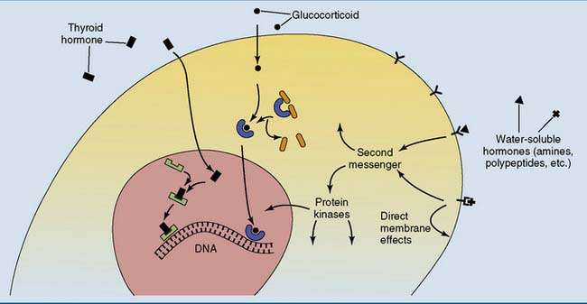
A key is useless without a matching lock, and a hormone or other extracellular signaling molecule is useless without a matching receptor in its target cell. The receptor is an allosteric protein that changes its conformation when it binds the signaling molecule. Messengers that do not enter the cell activate receptors in the plasma membrane, and many of those that can enter activate receptors in the cytoplasm or nucleus (Fig. 17.1).


Receptor binding triggers intracellular signaling cascades with protein-protein interactions and enzymatic reactions. The phosphorylation of cellular proteins by protein kinases is a recurrent feature of hormonally induced signaling cascades. These cascades regulate metabolic enzymes, membrane transporters, ion channels, and genes. This chapter describes the most important receptor mechanisms and signaling cascades.


Receptor-hormone interactions are noncovalent, reversible, and saturable


Like the binding of a substrate to its enzyme (see Chapter 4) or an antigen to its antibody (see Chapter 15), hormone-receptor binding is always noncovalent. Being noncovalent, it is reversible. The receptor-hormone complex (RċH) can easily dissociate back into free receptor (R) and free hormone (H):


image


The dissociation constant KD of the receptor-hormone complex is defined as


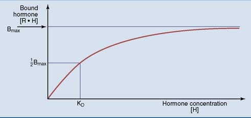
KD corresponds to the hormone concentration [H] at which half the receptor molecules are converted to the receptor-hormone complex. It describes the affinity between hormone and receptor.


Hormone binding shows saturation kinetics (Fig. 17.2). At a hormone concentration far above KD, almost all receptors are occupied. The physiological response is near maximal and cannot be augmented by adding even more hormone. This is equivalent to zero-order kinetics for enzymes. Maximal binding (Bmax) corresponds to the number of receptor molecules in the cell.


Many neurotransmitter receptors are ion channels


The job of a neurotransmitter is to change the membrane potential of the postsynaptic cell and to do it quickly. Rather than triggering lengthy signaling cascades, the transmitter should act as directly as possible on the ion channels that determine the membrane potential. The fastest and most direct mechanism is binding of the neurotransmitter to a ligand-gated ion channel in the plasma membrane.


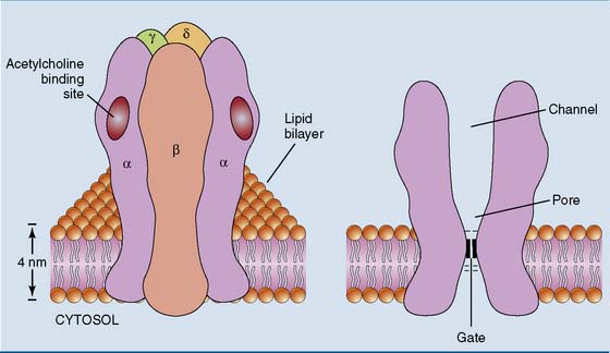
The nicotinic acetylcholine receptor in the neuromuscular junction is a classic example. This receptor is a channel for the monovalent cations sodium and potassium, with five subunits that each contribute to the channel (Fig. 17.3). The channel is closed in the resting state, opening only when acetylcholine binds. Opening of the channel causes a rapid influx of sodium down its electrochemical gradient, which depolarizes the membrane.


There is a whole family of ligand-gated ion channels. They all consist of five subunits but differ in their ligand-binding specificities and ionic selectivities. Most excitatory neurotransmitters open sodium channels, and inhibitory neurotransmitters open chloride or potassium channels.


The ligand-gated ion channels come in many different variants. For example, the subunits of the nicotinic acetylcholine receptors in the brain are slightly different from those of the receptor in the neuromuscular junction. Nicotine stimulates nicotinic receptors in the brain but not in the neuromuscular junction, and the arrow poison curare blocks nicotinic receptors in the neuromuscular junction but not in the brain. A drug that activates a receptor is called an agonist, and a drug that blocks a receptor is called an antagonist. Like enzymes, receptors are subject to competitive, noncompetitive, and irreversible inhibition by drugs and toxins.


Receptors for steroid and thyroid hormones are transcription factors


Unlike neurotransmitters, the steroid and thyroid hormones can diffuse across membranes and activate receptors in the cytoplasm or the nucleus. For example, the glucocorticoid receptor resides in the cytoplasm, complexed to cytoplasmic proteins that are released when the hormone binds.


The hormone-receptor complex translocates to the nucleus, where it binds to hormone response elements (HREs) in the promoters and enhancers of genes. Approximately 1% of all genes have glucocorticoid response elements in their regulatory sites. This implies two levels of targeting: Only cells that possess the receptor can respond to the hormone, and, within the cell, only genes that possess the appropriate response element are regulated by the hormone.


The receptors for steroid hormones, thyroid hormones, retinoic acid, and calcitriol all belong to the same superfamily of hormone-regulated transcription factors. All are zinc finger proteins that bind their response elements in a dimeric form, although the details are variable. For example, unstimulated thyroid hormone receptors are located in the nucleus rather than the cytoplasm.


An inherited deficiency of a receptor makes the cells unable to respond to the matching hormone (Clinical Examples 17.1 and 17.2).


Seven-transmembrane receptors are coupled to G proteins


Being unable to enter their target cells, water-soluble hormones deliver their message at the cell surface. Their receptors are integral membrane proteins with three functional domains. The extracellular domain binds the hormone; one or several transmembrane α-helices penetrate the lipid bilayer; and the intracellular domain is coupled with an effector mechanism.


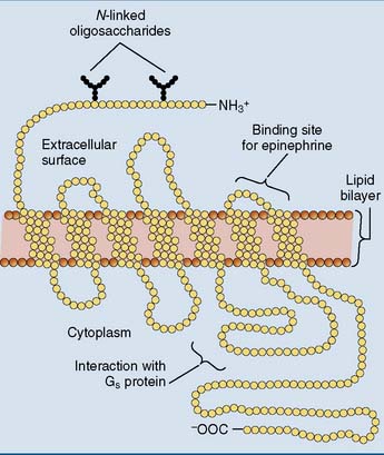
Most hormone receptors belong to a family of membrane proteins that crisscross the membrane seven times (Fig. 17.4). These receptors do not form a channel and possess no enzymatic activities, but they trigger their signaling cascades by activating a guanine nucleotide-binding G protein.


The G protein is attached to the cytoplasmic surface of the plasma membrane. Its three subunits are designated α (molecular weight [MW] 45,000), β (MW 35,000), and γ (MW 7000). The α subunit has a nucleotide binding site that can accommodate either guanosine diphosphate (GDP) or guanosine triphosphate (GTP). β and γ subunits function as a single unit, but the α subunit is only loosely associated with βγ.


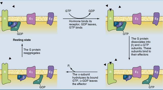
The function of the G protein is described in Figure 17.5. The inactive G protein is associated with the unstimulated receptor, with GDP bound to the α subunit. Hormone binding changes the conformation of the receptor and the attached G protein. As a result, the α subunit of the G protein loses its affinity for GDP, which dissociates away and is replaced by GTP.


Once GTP is bound, the G protein leaves the receptor and breaks up into the α-GTP subunit and the βγ complex. Both α-GTP and βγ diffuse along the inner surface of the plasma membrane, where they bind to target proteins known as effectors. The components of the activated G protein are membrane-bound messengers that transmit a signal from the receptor to the effector. In some cases, α-GTP and βγ can activate their effectors without fully separating from each other.


The α subunit possesses a GTPase activity that is stimulated by its interaction with the effector. As a result, the α subunit quickly hydrolyzes its bound GTP to GDP and inorganic phosphate. GDP remains bound to the α subunit, but the α-GDP complex no longer acts on the effector. Rather than transmitting a signal, it returns to the βγ complex. All G proteins exist in two forms: an active GTP-bound form that acts on the effector, and an inactive GDP-bound form that does not.


Several molecular forms of α, β, and γ subunits are expressed in different cells. G proteins are classified according to the structure and function of their α subunit. For example, the α-GTP complex of the Gs proteins stimulates adenylate cyclase, and the α-GTP complex of the Gi proteins inhibits adenylate cyclase.


The βγ complex transmits signals as well. The myocardium, for example, responds to acetylcholine from the vagus nerve through a receptor that couples to a Gi protein. The βγ complex of this Gi protein opens a potassium channel in the membrane, thereby hyperpolarizing the membrane and slowing down the heart.


Adenylate cyclase is regulated by G proteins


Hormone-activated G proteins carry messages along the plasma membrane but do not travel across the cytoplasm. To send a signal into the interior of the cell, the G protein induces the synthesis of a small, diffusible molecule known as a second messenger.


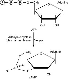
The second messenger cyclic adenosine monophosphate (cAMP) is synthesized by adenylate cyclase in the plasma membrane:


The rapid hydrolysis of pyrophosphate (PPi) by cellular pyrophosphatases makes this reaction irreversible. Adenylate cyclases are integral membrane proteins that are stimulated by the αs subunit of the stimulatory G proteins (Gs proteins). Humans have nine isoenzymes of adenylate cyclase that are encoded by different genes, are expressed in different cell types, and have different regulatory properties.


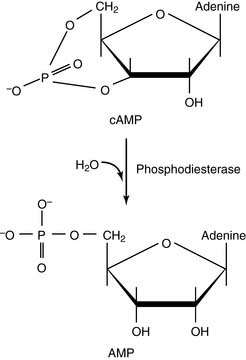
cAMP is degraded by phosphodiesterases:


There are 11 families of phosphodiesterases for the inactivation of cAMP and the related second messenger cyclic guanosine monophosphate (cGMP), with at least 100 different molecular forms. Most are inhibited by methylxanthines, including caffeine, theophylline, and aminophylline. These drugs potentiate the effects of cAMP. After drinking a cup of coffee, for example, the level of plasma free fatty acids rises because fat breakdown in adipose tissue is stimulated by cAMP.


Hormones can both activate and inhibit the cAMP cascade


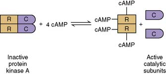
The most important target of cAMP is protein kinase A. In the absence of cAMP, two catalytic subunits of this enzyme form an inactive complex with two regulatory subunits. When the cAMP level rises, the two regulatory subunits bind up to four cAMP molecules, and the active catalytic subunits are released:


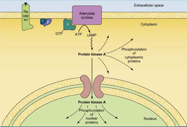
The catalytic subunits phosphorylate proteins on serine and threonine side chains. The enzyme phosphorylates only a small proportion of the cellular proteins, including several metabolic enzymes and nuclear transcription factors. In addition, many of the actions of protein kinase A are localized to specific intracellular sites because a portion of the enzyme is already bound to A-kinase anchoring proteins (AKAPs), which place the enzyme near its substrate proteins.


The cAMP cascade amplifies the hormonal signal (Fig. 17.6). For example, the binding of a single epinephrine molecule to a β-adrenergic receptor activates up to 20 Gs proteins. Each αs-GTP subunit activates adenylate cyclase long enough to cause the synthesis of hundreds of cAMP molecules. Although only four cAMP molecules are needed to activate two catalytic subunits of protein kinase A, each active subunit phosphorylates hundreds or thousands of proteins before it returns to the regulatory subunits.


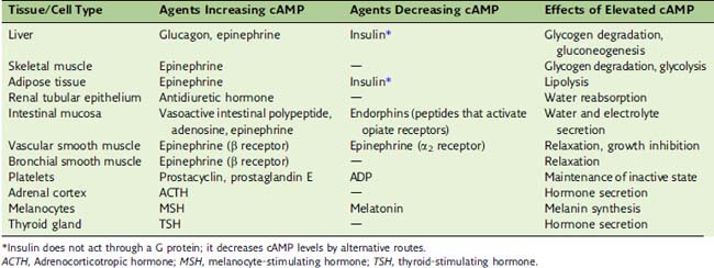
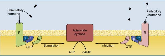
Some hormones do not stimulate but rather inhibit adenylate cyclase (see Table 17.1). In most cases inhibition is mediated by the αi subunit of an inhibitory G protein (Gi). Most cells possess both Gs-linked and Gi-linked hormone receptors, and the activity of adenylate cyclase depends on the balance between stimulatory and inhibitory hormones (Fig. 17.7).


Many hormones and neurotransmitters can act through different receptors and second messengers. For example, epinephrine (adrenaline) can raise the cAMP level by activating β-adrenergic receptors, or it can reduce cAMP by activating α2-adrenergic receptors. A third receptor type, the α1-adrenergic receptor, raises the calcium level by activating the IP3 second messenger system (see section “Phospholipase C generates two second messengers”). Therefore the effect of epinephrine depends on the type of receptor that is present on the cell. Acetylcholine acts not only on nicotinic receptors, which are ligand-gated ion channels, but also on several subtypes of muscarinic receptors, which are linked to G proteins and either lower the cAMP level or raise the levels of IP3 and calcium.


Jun 18, 2016 | Posted by in BIOCHEMISTRY | Comments Off on Intracellular Messengers

Full access? Get Clinical Tree

Get Clinical Tree app for offline access