Integration of metabolism in the whole body

chapter 18 Integration of metabolism in the whole body


KEY POINTS


The need for coordination
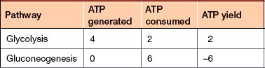

The previous two chapters outline the way in which the reactions in a cell are organised into discrete pathways, and explain that these pathways can be anabolic, catabolic or amphibolic. For every anabolic pathway described there is an equivalent, opposing, catabolic pathway.


It is imperative that the body is able to control the relative rates of flux through pathways for a number of reasons:


This chapter focuses on the way in which the organs of the body alter their metabolism to cope with changes in the availability of fuel sources, in particular glucose, and the way in which this is coordinated by the actions of the hormones insulin and glucagon.


Balancing blood glucose
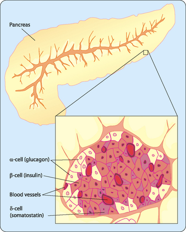

Glucose circulating in the blood provides a means by which fuel can be provided to cells, tissues and organs that require it. However, the concentration of glucose in the blood changes continuously. After eating, the concentration will rise as the processes of digestion and absorption add more glucose to the bloodstream. Conversely, if no new glucose is available from food then, as cells consume glucose as fuel, blood glucose concentrations ([BG]) will fall.


[BG] is strictly controlled in the human body and is maintained at 4–7 mM; hypoglycaemia (low [BG]) and hyperglycaemia (high [BG]) are both detrimental to the body.


Hypoglycaemia is a problem because, while some tissues can use alternative fuel sources such as fatty acids or amino acids, the brain and red blood cells have an absolute requirement for at least some glucose as fuel. If [BG] falls too low, the brain will start to shut down as it runs out of an adequate fuel supply, leading to coma and even death.


Hyperglycaemia is also problematic. Normally the kidneys will retain all of the glucose in the blood. However, if [BG] is raised above the renal threshold for glucose then they are no longer able to stop glucose being lost to the urine. This is detrimental in two ways; first, valuable fuel is lost from the body, and second, as the concentration of glucose increases in the urine more water is required to keep it soluble and this leads to more frequent urination and subsequent dehydration.


Humans do not eat all of the time and therefore the body has to cope with a large intake of fuel when food is consumed, more than is required for our energy needs, and periods between meals when glucose is no longer available from the intestine. To do this, the body controls key metabolic pathways to either store energy or access stored energy to make sure that [BG] is maintained.


These pathways are present in the insulin-sensitive tissues: liver, kidney, adipose tissue and skeletal muscle. Not all pathways are present in all of these tissues but together they can coordinate to maintain [BG].


The pancreas


The pancreas is a secretory organ that lies posterior to the stomach (Fig 18-1) and contains both an exocrine portion, which secretes digestive enzymes, and an endocrine portion, known as islets of Langerhans, which secrete insulin, glucagon and other hormones. The exocrine portion is made up of groups of cells known as acini. Digestive enzymes are secreted into the pancreatic duct, which empties into the gastrointestinal tract (Table 15-2). These enzymes catalyse the catabolism of carbohydrates, lipids and proteins.


Stay updated, free articles. Join our Telegram channel

Jun 11, 2016 | Posted by in BIOCHEMISTRY | Comments Off on Integration of metabolism in the whole body

Full access? Get Clinical Tree

Get Clinical Tree app for offline access