Insertion of Inferior Vena Cava Filters
Parth B. Amin
Joss D. Fernandez
Inferior vena cava filters have supplanted surgical interruption of the vena cava in the prevention of venous thrombotic embolic events. The ease of implantation has resulted in an increase and varied use despite very specific role they play in the management of patients with venous thromboembolic (VTE) disease. At present, the only absolute indications for placement include patient populations with (1) the development of a pulmonary embolus (PE) while on therapeutic anticoagulation and (2) an absolute contraindication to anticoagulation in patients with deep venous thrombosis (DVT). Relative indications include prophylaxis in patient who cannot be anticoagulated but are at high risk for DVT and those patients who have had a PE with continued residual DVT and may not tolerate another PE event. It is important to note that IVC filters may reduce episodes of pulmonary embolism, but have not been shown to reduce mortality. Long term, IVC filters are associated with an increased risk of lower extremity venous insufficiency. Other complications of IVC filters include migration, perforation of the vena cava or bowel, and complete thrombosis.
The two preferred routes of access are a right transfemoral venous approach and right transjugular venous approach. Alternative access sites include the left femoral and jugular veins and the left or right subclavian veins. Fluoroscopic guidance is the primary method used and will be the primary focus of this chapter. Both transabdominal and intravascular ultrasound have been used to guide successful placement of IVC filters. A brief discussion of suprarenal filters, congenital venous anomalies, and other special situations are also included in this text.
SCORE™, the Surgical Council on Resident Education, classified insertion of inferior vena cava filter as an “ESSENTIAL COMMON” procedure.
STEPS IN PROCEDURE
Choose approach
Transfemoral approach versus internal jugular approach
Access the vein percutaneously and dilate it
Perform cavogram
Measure IVC diameter
Identify renal veins
Deploy device
Withdraw wires and delivery device
HALLMARK ANATOMIC COMPLICATIONS
Femoral arterial puncture
Carotid arterial puncture
Groin hematoma
Neck hematoma
Pneumothorax
Hemothorax
Deep venous thrombosis
Iliac vein perforation
IVC thrombosis
Renal vein thrombosis
Filter migration
Filter erosion
LIST OF STRUCTURES
Anterior superior iliac spine
Pubic tubercle
Inguinal ligament
Femoral artery
Superficial femoral artery
Profunda femoris artery
Femoral vein
Right and left iliac veins
Inferior vena cava
Hepatic veins
Right atrium
Right and left renal veins
Lumbar veins
Carotid artery
Sternocleidomastoid muscle
Sternal head
Clavicular head
Subclavian vein
Superior vena cava
Innominate vein
Placement of Inferior Vena Cava Filter (Fig. 114.1)
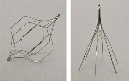
Multiple devices have been approved by the Food and Drug Administration for prevention of VTE disease (Table 114.1). Some examples are shown in Figure 114.1. Although the specific mechanisms by which different manufacturers design deployment devices varies, the basic principles of placement are similar. Familiarize yourself with the device that you plan to use.
A right transjugular approach or right transfemoral approach is most often selected. Identification of anatomic landmarks is paramount and can be facilitated by ultrasound in difficult situations.
Femoral Vein Approach (Fig. 114.2)
Local anesthetic is administered in the skin overlying the planned puncture site. Initial percutaneous access is then performed. This can be done using a standard Seldinger-type needle. A Micropuncture introducer set (Cook Incorporated, Bloomington, Indiana) is often used for initial percutaneous entry. This allows for the placement of a low-profile 0.018-inch wire into a 21-gauge Seldinger-type needle. Exchange can then be performed for a 4-French coaxial catheter, which allows for a 0.035-inch wire to be placed into the vena cava. Inadvertent arterial puncture is better tolerated with this system than a standard 18-gauge needle, although in experienced hands, complication rates are low with both methods even when patients are fully anticoagulated.
 Figure 114.1 Representative types of vena cava filter devices (from Fischer’s Mastery of Surgery, with permission). |
Table 114.1 Characteristics of IVC Filter Devices Available in the United States | |||||||||||||||||||||||||||||||||||||||||||||||||||||||||||||||||||||||||||||||||||||||||||||||||||||||||||||||||||||||||||||||||||||||||||||||||||||||||||||||||||||||
|---|---|---|---|---|---|---|---|---|---|---|---|---|---|---|---|---|---|---|---|---|---|---|---|---|---|---|---|---|---|---|---|---|---|---|---|---|---|---|---|---|---|---|---|---|---|---|---|---|---|---|---|---|---|---|---|---|---|---|---|---|---|---|---|---|---|---|---|---|---|---|---|---|---|---|---|---|---|---|---|---|---|---|---|---|---|---|---|---|---|---|---|---|---|---|---|---|---|---|---|---|---|---|---|---|---|---|---|---|---|---|---|---|---|---|---|---|---|---|---|---|---|---|---|---|---|---|---|---|---|---|---|---|---|---|---|---|---|---|---|---|---|---|---|---|---|---|---|---|---|---|---|---|---|---|---|---|---|---|---|---|---|---|---|---|---|---|---|
| |||||||||||||||||||||||||||||||||||||||||||||||||||||||||||||||||||||||||||||||||||||||||||||||||||||||||||||||||||||||||||||||||||||||||||||||||||||||||||||||||||||||
Stay updated, free articles. Join our Telegram channel

Full access? Get Clinical Tree


