CHAPTER 31 Infratemporal and pterygopalatine fossae and temporomandibular joint
INFRATEMPORAL FOSSA
The infratemporal fossa lies deep to the ramus of the mandible. It communicates with the temporal fossa deep to the zygomatic arch and the pterygopalatine fossa through the pterygomaxillary fissure. The major structures that occupy the infratemporal fossa are the lateral and medial pterygoid muscles, the mandibular division of the trigeminal nerve, the chorda tympani branch of the facial nerve, the otic parasympathetic ganglion, the maxillary artery and the pterygoid venous plexus.
The infratemporal fossa has a roof, and lateral and medial walls, and is open to the neck posteroinferiorly, i.e. the fossa has no anatomical floor. The roof is formed by the infratemporal surfaces of the temporal bone and of the greater wing of the sphenoid, and contains the foramena ovale and spinosum and the petrotympanic fissure: it is open superiorly to the temporal fossa. The medial wall is formed anteriorly by the lateral pterygoid plate of the pterygoid process of the sphenoid, and more postero-medially by the pharynx and tensor and levator veli palatini. It contains the pterygomaxillary fissure across which structures pass between the infratemporal and pterygopalatine fossae (Fig. 31.1). The lateral wall is formed by the medial surface of the ramus of the mandible.
Fig. 31.1 A, Pterygopalatine fossa after detachment of the mandible and removal of the zygomatic arch; B, anteromedial view; C, lateral view; arrows show that the floor of the temporal fossa is open medially to the infratemporal fossa and laterally to the region containing the masseter.
(From Drake, Vogl and Mitchell 2005.)
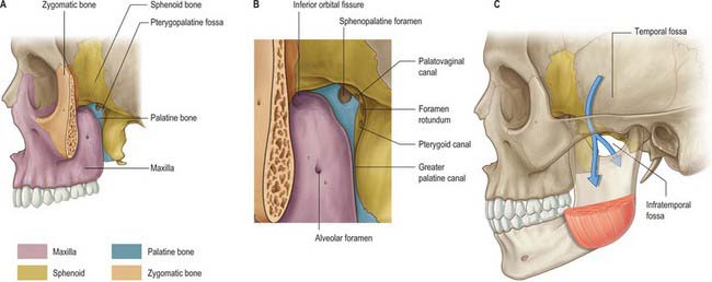
Lateral pterygoid provides a key to understanding the relationships of structures within the infratemporal fossa. This muscle lies in the roof of the fossa and runs anteroposteriorly in a more or less horizontal plane from the region of the pterygoid plates to the mandibular condyle (Fig. 31.1). Branches of the mandibular nerve and the main origin of medial pterygoid are deep relations and the maxillary artery is superficial. The buccal branch of the mandibular nerve passes between the two heads of lateral pterygoid. Medial pterygoid and the lingual and inferior alveolar nerves emerge below its inferior border and the deep temporal nerves and vessels emerge from its upper border. A venous network, the pterygoid venous plexus, lies around and within lateral pterygoid and is important in the spread of infection.
BONES
The sphenoid bone, the paired maxillae and temporal bones, and the mandible, collectively provide the skeletal framework to the infratemporal and pterygopalatine regions. The mandible and the two temporal bones articulate at the right and left temporomandibular joints. The disarticulated maxilla is described in Chapter 29, the temporal bone is described in Chapter 36, and the sphenoid and mandible are described here.
Sphenoid bone
The sphenoid bone lies in the base of the skull between the frontal, temporal and occipital bones. It has a central body, paired greater and lesser wings that spread laterally from the body, and two pterygoid processes that descend from the junction of the body and greater wings (Fig. 31.2).
Fig. 31.2 Sphenoid bone. A, anterior view; B, posterior view; C, superior view.
(From Sobotta 2006.)
Body
The body of the sphenoid is cuboidal. It contains two air sinuses, separated by a septum (see Ch. 32). Its cerebral (superior) surface articulates in front with the cribriform plate of the ethmoid bone. Anteriorly is the smooth jugum sphenoidale, which is related to the gyri recti and olfactory tracts. The jugum is bounded behind by the anterior border of the sulcus chiasmatis, which leads laterally to the optic canals. Posteriorly is the tuberculum sellae, behind which is the deeply concave sella turcica. In life the sella contains the hypophysis cerebri in the hypophysial fossa. Its anterior edge is completed laterally by two middle clinoid processes, while posteriorly the sella turcica is bounded by a square dorsum sellae, the superior angles of which bear variable posterior clinoid processes. The diaphragma sella and the tentorium cerebelli are attached to the clinoid processes (see Ch. 27). On each side, below the dorsum sellae, a small petrosal process articulates with the apex of the petrous part of the temporal bone. The body of the sphenoid slopes directly into the basilar part of the occipital bone posterior to the dorsum sellae, together these bones form the clivus. In the growing child this is the site of the spheno-occipital synchondrosis: premature closure of this joint gives rise to the skull appearances seen in achondroplasia.
The lateral surfaces of the body are united with the greater wings and the medial pterygoid plates. A broad carotid sulcus accommodates both the internal carotid artery and the cranial nerves associated with the cavernous sinus above the root of each wing (see Ch. 27). The sulcus is deepest posteriorly. It is overhung medially by the petrosal part of the temporal bone and has a sharp lateral margin, the lingula, which continues back over the posterior opening of the pterygoid canal.
A median triangular, bilaminar sphenoidal crest on the anterior surface of the body of the sphenoid makes a small contribution to the nasal septum. The anterior border of the crest joins the perpendicular plate of the ethmoid bone, and a sphenoidal sinus opens on each side of it (see Ch. 32). In the articulated state the sphenoidal sinuses are closed anteroinferiorly by the sphenoidal conchae, which are largely destroyed when disarticulating a skull. Each half of the anterior surface of the body of the sphenoid possesses a superolateral depressed area joined to the ethmoid labyrinth which completes the posterior ethmoidal sinuses; a lateral margin which articulates with the orbital plate of the ethmoid above and the orbital process of the palatine bone below; and an inferomedial, smooth, triangular area, which forms the posterior nasal roof, and near whose superior angle lies the orifice of a sphenoidal sinus.
The inferior surface of the body of the sphenoid bears a median triangular sphenoidal rostrum, embraced above by the diverging lower margins of the sphenoidal crest. The narrow anterior end of the rostrum fits into a fissure between the anterior parts of the alae of the vomer, and the posterior ends of the sphenoidal conchae flank the rostrum, articulating with its alae. A thin vaginal process projects medially from the base of the medial pterygoid plate on each side of the posterior part of the rostrum, behind the apex of the sphenoidal concha.
Greater wings
The greater wings of the sphenoid curve broadly superolaterally from the body. Posteriorly each is triangular, fitting the angle between the petrous and squamous parts of the temporal bone at a sphenosquamosal suture. The cerebral surface contributes to the anterior part of the middle cranial fossa. Deeply concave, its undulating surface is adapted to the anterior gyri of the temporal lobe of the cerebral hemisphere. The foramen rotundum lies anteromedially and transmits the maxillary nerve. Posterolateral to the foramen rotundum is the foramen ovale, which transmits the mandibular nerve, accessory meningeal artery and sometimes the lesser petrosal nerve, although the latter nerve may have its own canaliculus innominatus medial to the foramen spinosum. A small emissary sphenoidal foramen, which transmits a small vein from the cavernous sinus, lies medial to the foramen ovale (on one or both sides) in approximately 40% of skulls. The foramen spinosum, which transmits the middle meningeal artery and meningeal branch of the mandibular nerve, lies behind the foramen ovale.
The lateral surface is vertically convex and divided by a transverse infratemporal crest into temporal (upper) and infratemporal (lower) surfaces. Temporalis is attached to the temporal surface. The infratemporal surface is directed downwards and, with the infratemporal crest, is the site of attachment of the upper fibres of lateral pterygoid. It contains the foramen ovale and foramen spinosum. The small downward projecting spine of the sphenoid lies posterior to the foramen spinosum; the sphenomandibular ligament is attached to its tip. The medial side of the spine bears a faint anteroinferior groove for the chorda tympani nerve and appears in the lateral wall of the sulcus for the pharyngotympanic (auditory) tube. Medial to the anterior end of the infratemporal crest, a ridge passes downwards to the front of the lateral pterygoid plate, thereby forming a posterior boundary of the pterygomaxillary fissure.
The quadrilateral orbital surface of the greater wing faces anteromedially, and forms the posterior part of the lateral wall of the orbit. It has a serrated upper edge which articulates with the orbital plate of the frontal bone, and a serrated lateral margin which articulates with the zygomatic bone. Its smooth inferior border is the posterolateral edge of the inferior orbital fissure, and its sharp medial margin forms the inferolateral edge of the superior orbital fissure, on which a small tubercle gives partial attachment to the common anular ocular tendon. Below the medial end of the superior orbital fissure a grooved area forms the posterior wall of the pterygopalatine fossa; the latter is pierced by the foramen rotundum.
The irregular margin of the greater wing, from the body of the sphenoid to the spine, is an anterior limit of the medial half of the foramen lacerum. It also displays the posterior aperture of the pterygoid canal. Its lateral half articulates with the petrous part of the temporal bone at a sphenopetrosal synchondrosis. Inferior to this, the sulcus tubae contains the cartilaginous pharyngotympanic (auditory) tube. Anterior to the spine of the sphenoid the concave squamosal margin is serrated – bevelled internally below, externally above – for articulation with the squamous part of the temporal bone. The tip of the greater wing, bevelled internally, articulates with the sphenoidal angle of the parietal bone at the pterion. Medial to this, a triangular rough area articulates with the frontal bone: its medial angle is continuous with the inferior boundary of the superior orbital fissure, and its anterior angle joins the zygomatic bone by a serrated articulation.
Lesser wings
The lesser wings of the sphenoid are triangular pointed plates that protrude laterally from the anterosuperior regions of the body. The superior surface of each wing is smooth and related to the frontal lobe of the cerebral hemisphere. The inferior surface is a posterior part of the orbital roof and upper boundary of the superior orbital fissure, and overhangs the middle cranial fossa. The posterior border projects into the lateral fissure of the cerebral hemisphere. The medial end of the lesser wing forms the anterior clinoid process. The anterior and middle clinoid processes are sometimes united to form a caroticoclinoid foramen. The lesser wing is connected to the body by a thin flat anterior root and a thick triangular posterior root, between which lies the optic canal. Growth of the posterior root is closely associated with variations in the geometry of the canal. The cranial opening of the canal may be duplicated, or more commonly, the division is incomplete.
Superior orbital fissure
The superior orbital fissure connects the cranial cavity with the orbit. It is bounded medially by the body of the sphenoid, above by the lesser wing of the sphenoid, below by the medial margin of the orbital surface of the greater wing, and laterally, between the greater and lesser wings, by the frontal bone. The contents of the superior orbital fissure are described in Chapter 39.
Pterygoid processes
The pterygoid processes descend perpendicularly from the junctions of the greater wings and body. Each consists of a medial and lateral plate, whose upper parts are fused anteriorly. The plates are separated below by the angular pterygoid fissure, whose margins articulate with the pyramidal process of the palatine bone, and diverge behind. Medial pterygoid and tensor veli palatini lie in the cuneiform pterygoid fossa between the plates. Above is the small, oval, shallow scaphoid fossa, which is formed by division of the upper posterior border of the medial plate. Part of tensor veli palatini is attached to the fossa. The anterior surface of the root of the pterygoid process is broad and triangular and forms the posterior wall of the pterygopalatine fossa: it is pierced by the anterior opening of the pterygoid canal.
Lateral pterygoid plate
The lateral pterygoid plate is broad, thin and everted. The lateral surface forms part of the medial wall of the infratemporal fossa; the lower part of lateral pterygoid is attached to it. The medial surface is the lateral wall of the pterygoid fossa; most of the deep head of medial pterygoid is attached to it. The upper part of its anterior border is a posterior boundary of the pterygomaxillary fissure, and the lower part articulates with the palatine bone. The posterior border is free.
Medial pterygoid plate
The medial pterygoid plate is narrower and longer than the lateral. Its lower end is continued into an unciform projection, the pterygoid hamulus, which curves laterally. The pterygomandibular raphe is attached to the hamulus and the tendon of tensor veli palatini winds around the hamulus. The lateral surface forms the medial wall of the pterygoid fossa and the medial surface provides a lateral boundary of the posterior nasal aperture. The medial plate is prolonged above on the inferior aspect of the body of the sphenoid as a thin vaginal process that articulates anteriorly with the sphenoidal process of the palatine bone and medially with the ala of the vomer. The plate articulates with the posterior border of the perpendicular plate of the palatine bone in the lower part of its anterior margin. Inferiorly it bears a furrow, which is converted anteriorly into the palatovaginal canal by the sphenoidal process of the palatine bone. The palatovaginal canal transmits pharyngeal branches of the maxillary artery and pterygopalatine ganglion. The pharyngobasilar fascia is attached to the whole of the posterior margin of the medial plate, and the superior pharyngeal constrictor is attached to its lower end. The small pterygoid tubercle is found at the upper end of the plate, just below the posterior opening of the pterygoid canal. The processus tubarius, which supports the cartilaginous pharyngeal end of the pharyngotympanic tube, projects back near the midpoint of the margin of the medial pterygoid plate.
Sphenoidal conchae
The sphenoidal conchae are two thin, curved small plates, attached anteroinferiorly to the body of the sphenoid bone. The superior concave surface of each forms the anterior wall and part of the floor of a sphenoidal sinus. In situ, each has vertical quadrilateral anterior and horizontal triangular posterior parts. The anterior part consists of a superolateral depressed area, which completes the posterior ethmoidal sinuses and joins below with the orbital process of a palatine bone, and a smooth and triangular inferomedial area, which forms part of the nasal roof and is perforated above by the round opening connecting the sphenoidal sinus and sphenoethmoidal recess.
Anterior parts of the two bones meet in the midline, and protrude as the sphenoidal crest. The horizontal part appears in the nasal roof and completes the sphenopalatine foramen. Its medial edge articulates with the rostrum of the sphenoid and the ala of the vomer. Its apex, directed posteriorly, is superomedial to the vaginal process of the medial pterygoid plate and joins the posterior part of the ala. A small conchal part sometimes appears in the medial wall of the orbit, lying between the orbital plate of the ethmoid in front, the orbital process of the palatine bone below and the frontal bone above.
Ossification
Until the seventh or eighth month in utero the sphenoid body has a presphenoidal part, anterior to the tuberculum sellae, with which the lesser wings are continuous, and a postsphenoidal part, consisting of the sella turcica and dorsum sellae, and integral with the greater wings and pterygoid processes. Much of the bone is preformed in cartilage. There are six ossification centres for the presphenoidal parts, and eight for the postsphenoidal parts.
Presphenoidal part
About the ninth week of fetal life, a centre appears in each wing, lateral to the optic canal, and a little later two bilateral centres appear in the presphenoidal body. Each sphenoidal concha has a centre, appearing superoposteriorly in the nasal capsule in the fifth month in utero. As this enlarges it partly surrounds a posterosuperior expansion of the nasal cavity, which becomes the sphenoidal sinus. The posterior conchal wall is absorbed and the sinus invades the presphenoid component. In the fourth year the concha fuses with the ethmoidal labyrinth and before puberty it fuses with the sphenoid and palatine bones. Its anterior deficiency persists as an opening for the sphenoidal sinus.
Postsphenoidal part
The first centres appear in the greater wings about the eighth week of fetal life, one in the basal cartilage of each wing below the foramen rotundum. These centres only contribute to the root of the greater wing (near the foramen rotundum and pterygoid canal). The remainder of the greater wing and the lateral pterygoid plate are ossified in mesenchyme. About the fourth month of fetal life two centres appear, flanking the sella turcica, and soon fuse. The medial pterygoid plates are also ossified in ‘membrane’, a centre in each probably appearing about the ninth or tenth week. The hamulus is chondrified during the third fetal month and at once begins to ossify. Medial and lateral pterygoid plates join about the sixth fetal month. During the fourth month, a centre appears for each lingula, soon joining the body. The optic canal in the neonate is relatively large and has a keyhole or ‘figure of eight’ shape rather than the circular profile seen in the adult.
Postnatal details
Presphenoidal and postsphenoidal parts fuse about the eighth month in utero, but an unciform cartilage persists after birth in lower parts of the junction. At birth the bone is tripartite and consists of a central part (body and lesser wings) and two lateral parts (each consisting of a greater wing and pterygoid process). During the first year the greater wings and body unite around the pterygoid canals and the lesser wings extend medially above the anterior part of the body, meeting to form the smooth, elevated jugum sphenoidale. By the 25th year, sphenoid and occipital bones are completely fused. An occasional vascular foramen, often erroneously termed the craniopharyngeal canal, is occasionally seen in the anterior part of the hypophysial fossa. Although the sphenoidal sinus can be identified in the fourth month of fetal life as an evagination of the posterior part of the nasal capsule, by birth it represents an outgrowth of the sphenoethmoidal recess. Pneumatization of the body of the sphenoid commences in the second or third year and spreads first into the presphenoid, and later invades the postsphenoid, part. It reaches full size in adolescence, but often enlarges further by absorption of its walls as age advances.
Certain parts of the sphenoid are connected by ligaments that may occasionally ossify, e.g. the pterygospinous ligament between the sphenoid spine and upper part of lateral pterygoid plate; the interclinoid ligament joining the anterior to the posterior clinoid process; and the caroticoclinoid ligament that connects the anterior to the middle clinoid process.
Premature synostosis of the junction between pre- and post-sphenoidal parts, or of the spheno-occipital suture, produces a characteristic appearance, obvious in profile, of an abnormal depression of the nasal bridge (hypertelorism).
Mandible
The mandible is the largest, strongest and lowest bone in the face. It has a horizontally curved body that is convex forwards, and two broad rami that ascend posteriorly (Fig. 31.3). The body of the mandible supports the mandibular teeth within the alveolar process. The rami bear the coronoid and condylar processes. Each condyle articulates with the adjacent temporal bone at the temporomandibular joint.
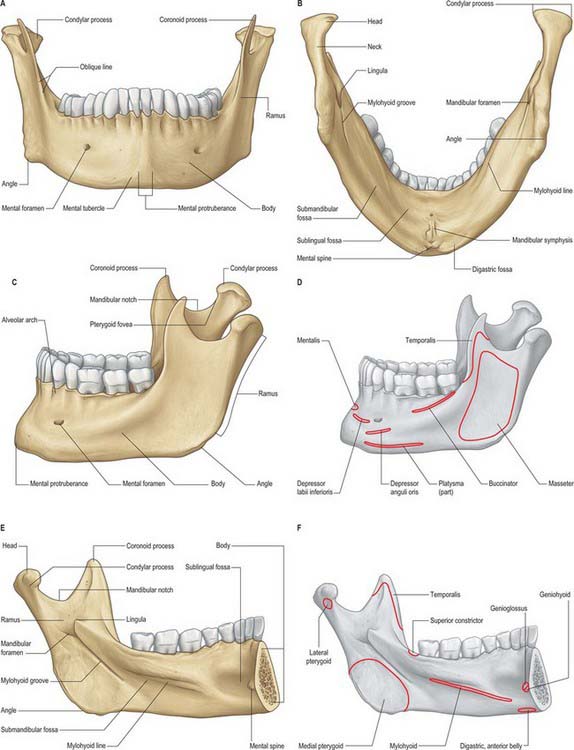
Fig. 31.3 Adult mandible. A, anterior view; B, inferior view; C, lateral view (with muscle attachments in D); E, internal view (with muscle attachments in F).
(Redrawn with permission from Sobotta 2006.)
Body
The body is somewhat U-shaped. It has external and internal surfaces separated by upper and lower borders. Anteriorly, the upper external surface shows an inconstant faint median ridge indicating the site of the fused symphysis menti. Inferiorly this ridge divides to enclose a triangular mental protuberance; its base is centrally depressed but raised on each side as a mental tubercle. The mental protuberance and mental tubercles constitute the chin. The mental foramen, from which the mental neurovascular bundle emerges, lies below either the interval between the premolar teeth, or the second premolar tooth, midway between the upper and lower borders of the body. The posterior border of the foramen is smooth, and accommodates the nerve as it emerges posterolaterally. A faint external oblique line ascends backwards from each mental tubercle, and sweeps below the mental foramen; it becomes more marked as it continues into the anterior border of the ramus.
The lower border of the body, the base, extends posterolaterally from the mandibular symphysis into the lower border of the ramus behind the third molar tooth. Near the midline on each side there is a rough digastric fossa which gives attachment to the anterior belly of digastric. Behind the fossa the base is thick and rounded: it has a slight anteroposterior convexity which changes to a gentle concavity as the ramus is approached, and so the base has an overall sinuous profile.
The upper border, the alveolar part, contains 16 alveoli for the roots of the lower teeth. It consists of buccal and lingual plates of bone joined by interdental and inter-radicular septa. Near the second and third molar teeth the external oblique line is superimposed upon the buccal plate. Like the maxilla, the form and depth of the tooth sockets is related to the morphology of the roots of the mandibular teeth. The sockets of the incisor, canine and premolar teeth usually contain a single root, while those for the three molar teeth each contain two or three roots. The third molar is variable in its position and root presentation. It may be impacted vertically, horizontally, mesially or distally, and its roots may be bulbous, hooked, divergent or convergent, and occasionally embrace the mandibular (inferior dental) canal (see Ch. 30). The internal surface of the mandible is divided by an oblique mylohyoid line that gives attachment to mylohyoid (and, above its posterior end, to the superior pharyngeal constrictor, some retromolar fascicles of buccinator, and the pterygomandibular raphe behind the third molar). The mylohyoid line extends from a point approximately 1 cm from the upper border behind the third molar as far forwards as the mental symphysis; it is sharp and distinct near the molars, but faint further forwards. The mylohyoid groove extends downwards and forwards from the ramus below the posterior part of the mylohyoid line and contains the mylohyoid neurovascular bundle. The area below the line is a slightly concave submandibular fossa and is related to the submandibular gland. The area above the line widens anteriorly into a triangular sublingual fossa and is related to the sublingual gland: the bone is covered by oral mucosa above the sublingual fossa as far back as the third molar. In an edentulous subject it may be necessary to reduce any ridge-like prominence of the mylohyoid line in order that dentures may fit without traumatizing the overlying oral mucosa.
Ramus
The mandibular ramus is quadrilateral, and has two surfaces (lateral and medial), four borders (superior, inferior, anterior and posterior) and two processes (coronoid and condylar). The lateral surface is relatively featureless and bears the (external) oblique ridge in its lower part. The medial surface presents, a little above centre, the mandibular foramen, through which the inferior alveolar neurovascular bundle passes to gain access to the mandibular canal (see below). Anteromedially, the mandibular foramen is overlapped by a thin, sharp triangular spine, the lingula, to which the sphenomandibular ligament is attached, and which is also the landmark for an inferior alveolar local anaesthetic block injection. Below and behind the foramen, the mylohyoid groove runs obliquely downward and forward.
The inferior border is continuous with the mandibular base and meets the posterior border at the angle, which is typically everted in males, but frequently inverted in females. The thin superior border bounds the mandibular incisure, which is surmounted in front by the somewhat triangular, flat, coronoid process and behind by the condylar process. The thick, rounded posterior border extends from the condyle to the angle, and is gently convex backwards above, and concave below. The anterior border is thin above, where it is continuous with the edge of the coronoid process, and thicker below where it is continuous with the external oblique line. The temporal crest is a ridge that descends on the medial side of the coronoid process from its tip to the bone just behind the third molar tooth. The triangular depression between the temporal crest and the anterior border of the ramus is the retromolar fossa.
The ramus and its processes provide attachment for the four primary muscles of mastication. Masseter is attached to the lateral surface, medial pterygoid is attached to the medial surface, temporalis is inserted into the coronoid process and lateral pterygoid is attached to the condyle.
Mandibular canal
The mandibular foramen leads into the mandibular canal, which runs obliquely downward and forward within the ramus, and then horizontally forward within the body under the roots of the molar teeth, with which it communicates by small openings. The canal is not always easy to define, especially anterior to the mental foramen. Its walls may be formed either by a thin layer of cortical bone or, more frequently, by trabecular bone. Although the buccal-lingual and superior-inferior positions of the canal vary considerably between mandibles, in general, the mandibular canal is situated nearer the lingual cortical plate in the posterior two-thirds of the bone, and closer to the labial cortical plate in the anterior third. Bilateral symmetry (location of the canal in each half of the mandible) is reported to be common. Near the mental foramen the inferior alveolar nerve branches into the mental nerve, which ultimately leaves the mandible via the mental foramen, and the incisive nerve, which remains within the bone and supplies the anterior teeth. The mental nerve may extend anteriorly for 2–3 mm within the mandible before curving back to the mental foramen (the ‘anterior loop’ of the mental nerve). The intra osseous anatomy of the mental nerve is of particular importance in dental implant surgery: the nerve may be damaged if the interforaminal area of the mandible is invaded during surgery or while harvesting block grafts from the symphysial region.
Coronoid process
The coronoid process projects upwards and slightly forwards as a triangular plate of bone. Its posterior border bounds the mandibular incisure, and its anterior border continues into that of the ramus.
Condylar process
The mandibular condyle varies considerably both in size and shape. When viewed from above, the condyle is roughly ovoid in outline, its anteroposterior dimension (approximately 1 cm) being roughly half its mediolateral dimension. The medial aspect of the condyle is wider than the lateral. The long axis of the condyle is not at right angles to the ramus, but diverges posteriorly from a strictly coronal plane. Thus, the lateral pole of the condyle lies slightly anterior to the medial, and if the long axes of the two condyles are extended, they meet at an obtuse angle (approximately 145°) at the anterior border of the foramen magnum. The articular head of the condyle joins the ramus through a thin bony projection, the neck of the condyle. The pterygoid fovea, a small depression situated on the anterior surface of the neck below the articular surface, receives part of the attachment of lateral pterygoid.
The condyle consists of a core of cancellous bone covered by a thin outer layer of compact bone whose intra-articular aspect is covered by layers of fibrocartilage. The condyle is the commonest site of mandibular fractures.
Accessory foramina of the mandible
These are usually unnamed and infrequently described, yet they are numerous. They may transmit auxiliary nerves to the teeth (from facial, mylohyoid, buccal, transverse cervical cutaneous and other nerves), and their occurrence is significant in dental anaesthetic blocking techniques.
Ossification
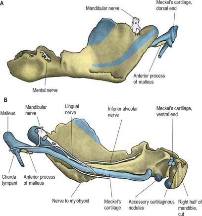
The mandible forms in dense fibromembranous tissue lateral to the inferior alveolar nerve and its incisive branch, and also in the lower parts of Meckel’s cartilage (first branchial arch). Each half is ossified from a centre that appears near the mental foramen about the sixth week in utero. From this site, ossification spreads medially and posterocranially to form the body and ramus, first below, and then around, the inferior alveolar nerve and its incisive branch. Ossification then spreads upwards, initially forming a trough, and later crypts, for the developing teeth. By the tenth week, Meckel’s cartilage below the incisor rudiments is surrounded and invaded by bone. Secondary cartilages appear later (Fig. 31.4): a conical mass, the condylar cartilage, extends from the mandibular head downwards and forwards in the ramus, and contributes to its growth in height. Although it is largely replaced by bone by midfetal life, its proximal end persists as proliferating cartilage under the fibrous articular lining until about the third decade. Another secondary cartilage, which soon ossifies, appears along the anterior coronoid border, and disappears before birth. One or two cartilaginous nodules also occur at the symphysis menti. At about the seventh month in utero these may ossify as variable mental ossicles in symphysial fibrous tissue: they unite with adjacent bone before the end of the first postnatal year.
Age changes in the mandible
At birth the two halves of the mandible are united by a fibrous symphysis menti (Fig. 31.5). The anterior ends of both rudiments are covered by cartilage, separated only by a symphysis. Until fusion occurs, new cells are added to each cartilage from symphysial fibrous tissue, and ossification on its mandibular side proceeds towards the midline. When the latter process overtakes the former, and ossification extends into median fibrous tissue, the symphysis fuses. At this stage the body is a mere shell which encloses the imperfectly separated sockets of deciduous teeth. The mandibular canal is near the lower border, and the mental foramen opens below the first deciduous molar and is directed forwards. The coronoid process projects above the condyle.
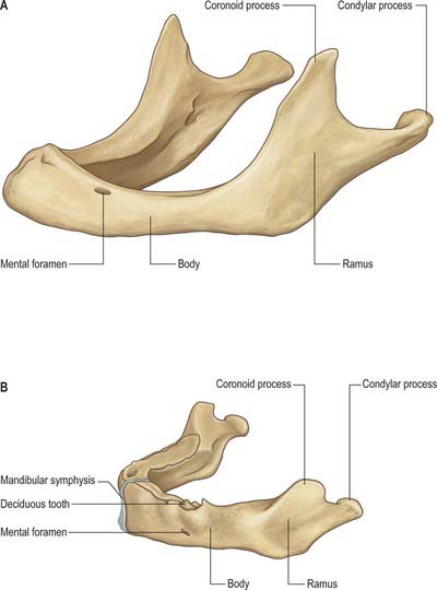
Fig. 31.5 A, Edentulous mandible; note position of mental foramen; B, Neonatal mandible.
(Redrawn with permission from Sobotta 2006.)
During the first three postnatal years, the two halves join at their symphysis from below upwards, although separation near the alveolar margin may persist into the second year. The body elongates, especially behind the mental foramen, providing space for three additional teeth. During the first and second years, as a chin develops, the mental foramen alters direction: it no longer faces forwards but now faces backwards, as in the adult mandible, and accommodates the changing direction of the emerging mental nerve.
In general terms, increase in height of the body of the mandible is achieved primarily by formation of alveolar bone associated with the developing and erupting teeth, although some bone is also deposited on the lower border. Increase in length of the mandible is accomplished by deposition of bone on the posterior surface of the ramus and concomitant compensatory resorption on the anterior surface (accompanied by deposition of bone on the posterior surface of the coronoid process and resorption on the anterior surface of the condylar process). Increase in width of the mandible is produced by deposition of bone on the outer surface of the mandible and resorption on the inner surface. An increase in the comparative size of the ramus compared with the body of the mandible occurs during postnatal growth and tooth eruption.
The role of the condylar cartilages in mandibular growth remains controversial. One view states that continued proliferation of this cartilage is primarily responsible for the increase in both the mandibular length and the height of the ramus. Alternatively, there is persuasive experimental evidence that proliferation of the condylar cartilage is an adaptive response to function, rather than being genetically determined. Condylar growth and remodeling has been shown to be influenced significantly by local factors, notably movement and loading of the temporomandibular joint, and to be relatively immune to systemic influences such as vitamin C and D deficiency. Considering the changes that occur in the dentition throughout life, continuous adaptation of the temporomandibular articulation is required in order to maintain functional occlusal alignment between the upper and lower arches of teeth: this adaptation is thought to be largely the result of ongoing condylar remodelling.
In adults, alveolar and subalveolar regions are about equal in depth, and the mental foramen appears midway between the upper and lower borders. If teeth are lost, alveolar bone is resorbed, which means that the mandibular canal (which runs parallel to the mylohyoid line) and the mental foramen come to lie much nearer to the superior border (Fig. 31.5), indeed, sometimes they may both disappear, so that the nerves lie just beneath the oral mucosa.
TEMPOROMANDIBULAR JOINT
The temporomandibular joint is a synovial joint between the articular fossa (also known as the mandibular fossa or glenoid fossa) of the temporal bone above and the mandibular condyle. It is unusual in that its articular surfaces are lined by fibro-cartilage (rather than hyaline cartilage) and its joint cavity is divided into two by an articular disc.
The articular eminence, a transversely elliptical region sinuously curved in the sagittal plane and tilted downwards anteriorly at approximately 25° to the occlusal plane, forms most of the articular surface of the mandibular fossa. Its steepness is variable, and it becomes flatter in the edentulous subject. Its anterior limit is the summit of the articular eminence, a transverse ridge that extends laterally out to the zygomatic arch as far as the articular tubercle. Articular tissue extends anteriorly beyond the articular summit and on to the preglenoid plane. Posteriorly it extends behind the depth of the fossa as far as the squamotympanic fissure. A postglenoid tubercle (at the root of the zygomatic arch, just anterior to the fissure) is usually poorly developed in human skulls.
The articular surface of the mandibular condyle is slightly curved and tilted forward at approximately 25° to the occlusal plane. Like the articular eminence, its slope is variable. In the coronal plane its shape varies from that of a gable (particularly marked in those whose diet is hard), to roughly horizontal in the edentulous.
It is probably impossible to measure the pressure developed on the articular surfaces of the human jaw joint when biting, however direct measurement of loads across the joint in animals has demonstrated significant intermittent loading during mastication. There is also irrefutable theoretical evidence based on Newtonian mechanics that the jaw joint is a weight-bearing joint. With a vertical bite force of 500 N on the left first molar, the right condyle must support a load of well over 300 N (Osborn 1995a). The non-working condyle is more loaded than the condyle on the working side, which may help explain why patients with a fractured condyle choose to bite on the side of the fracture.
Full access? Get Clinical Tree
