Infantile (Juvenile) Hemangioma
Jeremy C. Wallentine, MD
Key Facts
Clinical Issues
Most common tumor of infancy
Appear within 1st few weeks of life
Rapidly enlarge over several months
All eventually spontaneously regress
Microscopic Pathology
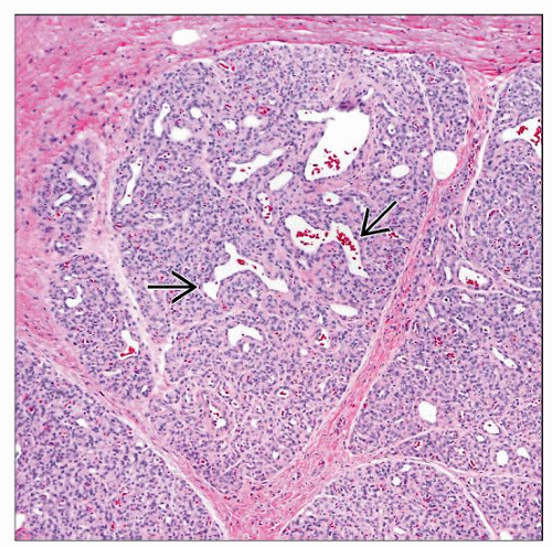
Multiple lobules composed of tightly packed small to moderate-sized capillaries
Lobules are separated by normal stroma
Plump endothelial cells that line small vascular spaces in early lesions
Progressive interstitial fibrosis in older lesions
Ancillary Tests
GLUT1 is most useful marker for diagnosis
Top Differential Diagnoses
Pyogenic granuloma
Arteriovenous malformation
Tufted angioma
 Clinical photograph of a medium-sized hemangioma in a female infant. Most of these lesions are located in the head and neck region. (Courtesy J. Hall, MD.) |
TERMINOLOGY
Synonyms
Hemangioma of infancy
Juvenile hemangioma
Cellular hemangioma of infancy
Strawberry nevus/hemangioma
Definitions
Vascular neoplasm of infancy with characteristic onset, rapid growth, and spontaneous involution
CLINICAL ISSUES
Epidemiology
Incidence
Most common tumor of infancy
Affects ˜ 4% of children
Gender
M < F
Ethnicity
Caucasians more frequently affected
Site


