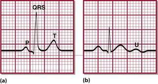
12 The factors affecting potassium balance have been described previously (p. 22). Hypokalaemia may be due to reduced potassium intake, but much more frequently results from increased losses or from redistribution of potassium into cells. As with hyperkalaemia, the clinical effects of hypokalaemia are seen in ‘excitable’ tissues like nerve and muscle. Symptoms include muscle weakness, hyporeflexia and cardiac arrhythmias. Figure 12.1 shows the changes that may be found on ECG in hypokalaemia. Fig 12.1 Typical ECG changes associated with hypokalaemia. (a) Normal ECG (lead II). (b) Patient with hypokalaemia: note flattened T-wave. U-waves are prominent in all leads.
Hypokalaemia

Diagnosis
Redistribution into cells
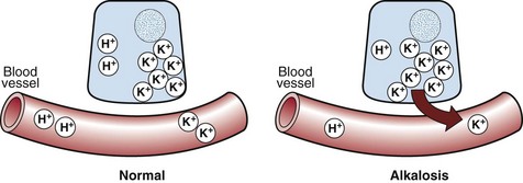
 Metabolic alkalosis. The reciprocal relationship between potassium and hydrogen ions means that in just the same way as metabolic acidosis is associated with hyperkalaemia, so metabolic alkalosis is associated with hypokalaemia. As the concentration of hydrogen ions decreases, so potassium ions move inside cells in order to maintain electrochemical neutrality (Fig 12.2).
 Treatment with insulin. Insulin stimulates cellular uptake of potassium, and plays a central role in treatment of severe hyperkalaemia (see pp. 22–23). It should come as no surprise therefore that when insulin is given in the treatment of diabetic ketoacidosis (see pp. 66–67), there is a risk of hypokalaemia. This is well recognized, and virtually all treatment protocols for diabetic ketoacidosis take this into account.
![]()
Stay updated, free articles. Join our Telegram channel

Full access? Get Clinical Tree


Hypokalaemia


