Hypofunction of the adrenal cortex

48


Hypofunction of the adrenal cortex


Adrenal insufficiency


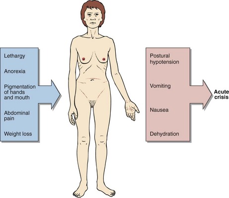
Acute adrenal insufficiency is a rare condition that if unrecognized is potentially fatal. Because of its low incidence it is often overlooked as a possible diagnosis. It is relatively simple to treat once the diagnosis has been made, and patients can lead a normal life. The clinical features of adrenal insufficiency are shown in Figure 48.1.


In areas where tuberculosis is endemic, adrenal gland destruction may be due to TB; in developed countries autoimmune disease is now the main cause of primary adrenal failure. Both cortisol and aldosterone production may be affected. Secondary failure to produce cortisol is more common. Frequently, this is due to long-standing suppression and subsequent impairment of the hypothalamic–pituitary–adrenocortical axis from therapeutic administration of corticosteroids. The causes of adrenal insufficiency are summarized in Figure 48.2.


Biochemical features


In addition to the clinical observations, a number of biochemical results may point towards adrenocortical insufficiency. These are hyponatraemia, hyperkalaemia and elevated serum urea, and may be seen in many patients with Addison’s disease.


In primary adrenal insufficiency, patients become hyponatraemic for two reasons. The lack of aldosterone leads to pathological sodium loss by the kidney that results in a contraction of the extracellular fluid volume, causing hypotension and pre-renal uraemia. Patients may develop life-threatening sodium depletion and potassium retention due to aldosterone deficiency. The hypovolaemia and hypotension stimulate AVP secretion, thus causing water retention. In the absence of cortisol, the kidneys’ ability to excrete a water load is impaired, thus exacerbating hyponatraemia. Overall, however, the total body water is reduced and this is reflected by the increase in the serum urea.


Lack of negative feedback of cortisol on the anterior pituitary results in an excessive secretion of ACTH. The structure of this hormone contains part of the amino acid sequence of melanocyte-stimulating hormone. Where excessive ACTH secretion occurs, patients may show darkening of the skin and mucous membranes.


Jun 18, 2016 | Posted by in BIOCHEMISTRY | Comments Off on Hypofunction of the adrenal cortex

Full access? Get Clinical Tree

Get Clinical Tree app for offline access