Chapter 1 How does an embryo form?
Human development begins when a spermatozoon fertilizes an oocyte. By definition, an embryo comprises the tissues formed once mitosis of an ovum (a fertilized oocyte) begins; thus even at the two-cell stage it is an embryo. These few cells multiply in number over an 8-week period into a fetus, by which time it will consist of many millions of cells. During the first two weeks (the pre-embryonic period) the embryo moves through the uterine tube to the uterus where it will implant. The differentiation that establishes the organ systems takes place between 3 and 8 weeks in the first 8 weeks following fertilization (the embryonic period). The period of time from the end of week 8 to full term (38 weeks) is a phase of growth and enlargement (the fetal period). The crucial phase during which there is potential for malformation is in the first 8 weeks, and this period is when the embryo is most vulnerable to environmental agents such as viruses and other teratogens. Table 1.1 summarizes the major events of the prenatal stages of development. The stages of the formation of an embryo are often described in relation to weeks of development.
Table 1.1 Stages of development before birth
| Time period | Stage | Main events |
|---|---|---|
| Conception to week 2 | Pre-embryonic period | |
| 2nd to 8th week | Embryonic period | |
| 9th week to birth | Fetal period |
 Clinical box
The 1st week—Fertilization and formation of the blastocyst
A fertilized ovum has a diploid number of chromosomes and once the second meiotic division has been completed, the stage of cleavage can begin. This consists of a series of rapid mitotic cell divisions in which the ovum divides over a period of about 3 days, resulting in the so-called 16-cell-stage embryo (Figs 1.1 and 1.2A). Each cell is known as a blastomere. After each cleavage division, whilst the number of cells increases, the size of each cell diminishes. The solid sphere of cells that forms is known as a morula, because it was thought to resemble a mulberry! Each of these new daughter cells is, at this stage, pluripotential. In other words, each daughter cell has the potential to differentiate into cells of any lineage.
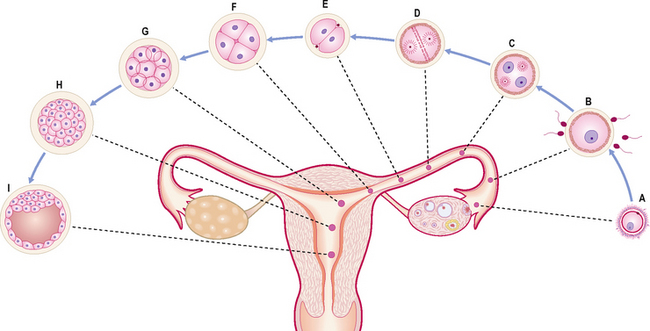
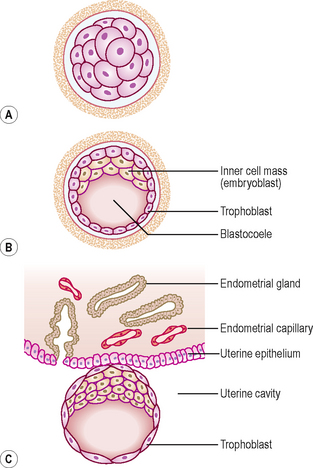
The morula soon shows signs of further differentiation. Cavities appear within the centre of the sphere of cells, forming a blastocyst, the cavity itself being the blastocoele (Fig. 1.2B, C). Once this stage has been reached the outer layer of the blastocyst soon thins to single-cell thickness to become the trophoblast, enclosing the enlarging fluid-filled blastocyst cavity. The central group of cells move to one pole of the blastocyst (the embryonic pole) to form the inner cell mass from which the whole embryo itself will form. The trophoblast contributes to the fetal component of the placenta (Fig. 1.3).

The process of morula and blastocyst formation occurs whilst the sphere of dividing cells is in transit along the uterine tube (Fig. 1.1). Fertilization takes place in the ampulla of the uterine tube, approximately 12–24 hours after ovulation. The first mitotic division of cleavage will be completed by the time that the two-cell stage embryo reaches the middle of the tube, at about 30 hours post-fertilization. By 3 days the morula of 12–16 cells will have reached the junction of the uterine tube and the uterus. By 4–5 days the fully formed blastocyst reaches the uterine lumen in preparation for implantation, which occurs a day later.
The 2nd week—Implantation and formation of bilaminar embryonic disc
At this stage the embryo is partly implanted in the endometrium. The implantation process initiates the decidual reaction or decidualization in the uterine stroma, the cells of which contribute the maternal component of the placenta. The trophoblast begins to differentiate: its inner part becomes a single layer of cells, hence its name the cytotrophoblast (Fig. 1.3A). The outer layer is more extensive and is the invasive layer. It is a syncytium, and at this stage, although it has invaded the endometrium, it has not invaded endometrial blood vessels. It is known as the syncytiotrophoblast. By this stage, the inner cell mass of the blastocyst has differentiated into two layers: the epiblast and the hypoblast (Fig. 1.3A). These two layers are in contact and form a bilaminar embryonic disc. Within the epiblast a cavity develops, the amniotic cavity, which fills with amniotic fluid. Some epiblast cells become specialized as amnioblasts, and they secrete the amniotic fluid. The exocoelomic membrane is derived from the hypoblast and lines the cavity that appears beneath the endoderm, the primary yolk sac (Fig. 1.3C). The fluid contained in this sac is the source of nutrition for the embryo before the placenta is fully formed and functional.
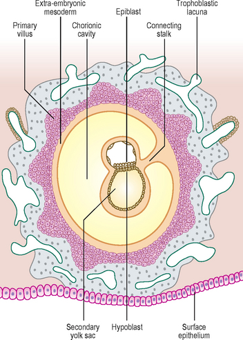
By 12 days there has been significant change particularly in the trophoblast. Small clefts appear in the syncytiotrophoblast called lacunae which communicate with the maternal endometrial sinusoids, thereby deriving nutritional support for the developing embryo (Fig. 1.4). Concurrent with this is the further development of the cytotrophoblast which is thickest at the embryonic pole of the conceptus. Clefts appear between the exocoelomic membrane and the cytotrophoblast (Fig. 1.4). These merge to form the extra-embryonic coelom and this cavity comes to almost completely surround the embryo. This is the chorionic cavity (Fig. 1.5).
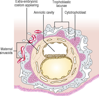
Fig. 1.4 Implanted blastocyst. At 12 days cavities develop which coalesce to form the extra-embryonic coelom.