Hobnail (Targetoid Hemosiderotic) Hemangioma
David Cassarino, MD, PhD
Key Facts
Terminology
Targetoid hemosiderotic hemangioma
Benign vascular proliferation, typically wedge-shaped, showing intravascular papillae and hobnailed endothelial cells
Etiology/Pathogenesis
Postulated to represent traumatized lymphangioma or hemangioma
Clinical Issues
Typically presents on lower extremities; also may occur on upper extremities, rarely in oral cavity
Microscopic Pathology
Superficial vessels are dilated and thin-walled
Deeper vessels are progressively smaller
Vessels are lined by small, bland-appearing endothelial cells with hobnail appearance
 Low-magnification view of a hobnail hemangioma demonstrates superficial dilated vascular spaces  in the papillary dermis with deeper small blood vessels and stromal hemosiderin deposits in the papillary dermis with deeper small blood vessels and stromal hemosiderin deposits  . . |
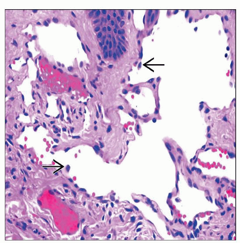 Higher power examination shows a hobnail hemangioma with superficial dilated vessels lined by small endothelial cells protruding into the lumina  and showing nuclear hyperchromasia. and showing nuclear hyperchromasia. |
TERMINOLOGY
Abbreviations
Hobnail hemangioma (HH)
Synonyms
Targetoid hemosiderotic hemangioma (clinical term)
Definitions
Benign vascular proliferation, typically wedge-shaped, showing intravascular papillae and hobnailed endothelial cells
ETIOLOGY/PATHOGENESIS
Unknown
Trauma has been implicated in some cases
Postulated to represent traumatized lymphangioma or hemangioma


