HIV-associated Collapsing Glomerulopathy
A. Brad Farris, III, MD
Key Facts
Clinical Issues
Up to 90% of cases occur in African-American blacks, mostly male
Rapid progression to ESRD
Hypertension uncommon (in < 50%)
Nephrotic-range proteinuria and ↑ creatinine
Treatment: HAART, ACE inhibitors
Macroscopic Features
Enlarged with cysts
Microscopic Pathology
Collapsing GS
Microcystic tubular dilatation (30-40% of cases)
Mononuclear cells (including lymphocytes and monocytes) and plasma cells
Interstitial inflammation and edema
Ancillary Tests
IF: IgM, C3, and C1q in capillary walls in segmental distribution
EM
Podocyte foot process effacement and multilamination of subepithelial GBM
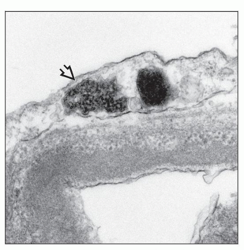
Tubuloreticular inclusions (TRIs) in endothelial cells, lymphocytes, and monocytes
Granular and, less commonly, granulofibrillar transformation of tubulointerstitial nuclei
Top Differential Diagnoses
Idiopathic collapsing glomerulopathy and FSGS
Diffuse mesangial hypercellularity and minimal change disease
TERMINOLOGY
Synonyms
HIV-associated nephropathy (HIVAN)
HIV-associated collapsing glomerulopathy
Definitions
Characteristic renal disease developing in setting of HIV infection
ETIOLOGY/PATHOGENESIS
Infectious Agents
HIV
Some suggest kidney is “reservoir” for HIV
HIV genome demonstrated in glomerular visceral and tubular epithelial cells in HIVAN
Abnormal maturation and differentiation of podocytes demonstrated (Barisoni et al)
Cytokines may have role
Abnormalities shown in transforming growth factor-β (TGF-β), basic fibroblast growth factor (bFGF), interleukin, and others
AIDS not required for condition to occur
Genetic Factors
Higher risk with African ancestry (except Ethiopian)
APOL1 polymorphism associated with HIVAN
Animal Models
Mice transgenic for HIV regulatory genes develop collapsing glomerulopathy
Renal HIV gene expression necessary for transgenic HIVAN
Normal kidneys fail to develop nephropathy when transplanted into HIV transgenic mice
Transplantation of transgenic kidneys into nontransgenic litter mates shows the renal HIVAN phenotype
CLINICAL ISSUES
Epidemiology
Incidence
Average incidence of acute kidney injury is 2.7 episodes per 100 person-years
Higher within 1st 3 months of disease recognition: 19.3 episodes per 100 person-years
Gender
Majority (˜ 70%) male, particularly according to original descriptions
Ethnicity
Historically, up to 90% of African American blacks
Rare in whites
Presentation
Proteinuria, nephrotic range
Severe, averaging 6-7 g/d
Edema, hypoalbuminemia, and hypercholesterolemia (nephrotic syndrome) uncommon
Renal dysfunction
Creatinine at diagnosis typically ˜ 5 mg/dL
Hypertension
Uncommon (in < 50%)
Microhematuria
Laboratory Tests
CD4 count does not correlate with HIVAN development
Hypercholesterolemia uncommon, possibly due to ↓ hepatic lipoprotein synthesis in AIDS patients
Treatment
Prognosis
Rapidly progressive, with ESRD often developing within months in most
Early in AIDS epidemic, mean time to dialysis < 2 months and median survival time of renal disease to death is 4.5 months
IMAGE FINDINGS
Ultrasonographic Findings
Enlarged, highly echogenic kidneys
MACROSCOPIC FEATURES
General Features
Pale cortex
0.5-1.0 mm cysts in cortex or at corticomedullary junction
Size
Enlarged, mean combined weight up to 500 g in adults
1.2-1.3x normal weight in children
MICROSCOPIC PATHOLOGY
Histologic Features
Glomeruli
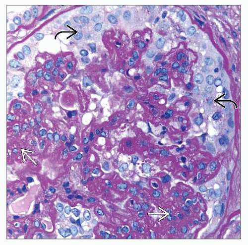
Collapsing glomerulopathy
Capillary loops globally or segmentally collapsed without corresponding mesangial matrix ↑
Shrunken down, global glomerulosclerosis (GS) ± cystic Bowman space dilatation, which contains proteinaceous material
± marked podocyte hyperplasia, protein resorption droplets, and mitoses (contrary to prior thought that podocytes could not undergo mitosis)
± enlarged glomeruli
Global GS reduces glomerulus to acellular sclerotic ball
Podocytes prominent
May simulate a crescent
Starts with hypertrophy and hyperplasia of visceral epithelial cells
Tubules
Microcystic tubular dilatation (30-40% of cases)
± scalloped tubular outline
± PAS(+) proteinaceous casts (fuchsin[+] on trichrome) in renal tubules
Autopsy studies have shown near-complete replacement of renal cortical parenchyma by microcysts
Patchy epithelial injury, regeneration, degeneration, and necrosis, and eventual tubular atrophy
Flattened tubular epithelium and mitotic figures
Tubular resorption droplets prominent
Interstitium
Interstitial inflammation and edema
Mononuclear cells (including lymphocytes and monocytes) and plasma cells
Interstitial fibrosis typically present
Vessels
Typically unremarkable
Rare cases of thrombotic microangiopathy
ANCILLARY TESTS
Immunofluorescence
Segmental IgM, C3, and C1q
± IgM and C3 granular positivity
Resorption droplets in podocytes and proximal tubules stain for multiple plasma proteins
Prominent immune complex deposit would be indicative of additional diagnosis
Hepatitis C virus (HCV), lupus-like GN, IgA nephropathy can all be seen in HIV
Electron Microscopy
Glomeruli
Podocytes enlarged with ≥ 1 dense, round 2° lysosome and enlarged vacuoles
Podocyte foot processes usually completely effaced
Stay updated, free articles. Join our Telegram channel

Full access? Get Clinical Tree


