, Tsunehisa Kaku2, Toru Sugiyama3 and Steven G. Silverberg4
(1)
Matsue City Hospital, Matsue, Shimane, Japan
(2)
Department of Health Sciences, Department of Health Sciences Graduate School of Medical Sciences, Fukuoka, Fukuoka, Japan
(3)
Department of Obstetrics and Gynecology, Iwate Medical University School of Medicine, Morioka, Iwate, Japan
(4)
Department of Pathology, University of Maryland School of Medicine, Baltimore, MD, USA
Oxyphilic differentiation is seen at least focally in at least half of ovarian CCCs, is prominent in 25–30 %, and may be the dominant (or occasionally the only) cell type in 5 % or less. The oxyphil cells, as in other tumors and other organs, are characterized by large quantities of amphophilic cytoplasm, often with enlarged atypical nuclei, and generally with no mitotic activity (Figs. 4.1, 4.2, 4.3, 4.4, 4.5, 4.6, 4.7, 4.8, 4.9, 4.10, 4.11, 4.12, and 4.13). These cells may be oncocytes, but they have never been shown to be packed with mitochondria like true oncocytes. In many instances, clear cells are seen adjacent to oxyphil cells, and some cells even have admixtures of clear and oxyphilic cytoplasm (Figs. 4.8, 4.9, 4.10, 4.11, 4.12, and 4.13).
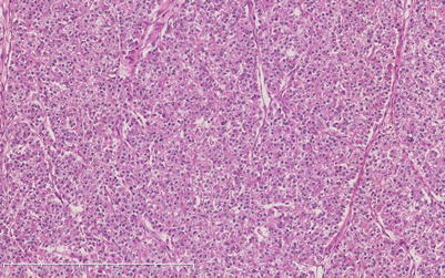
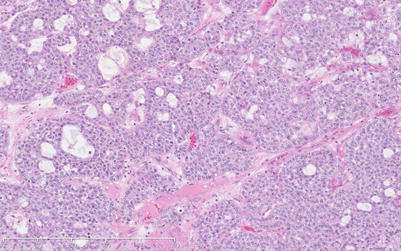
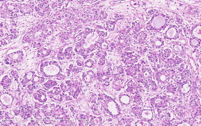
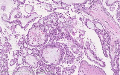
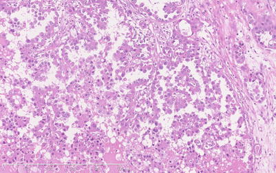
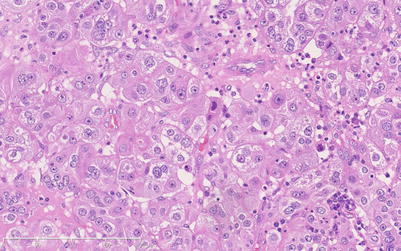
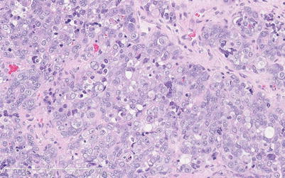
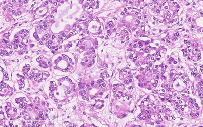
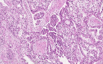
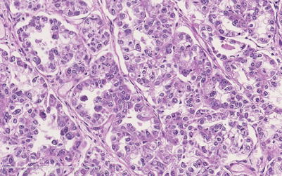
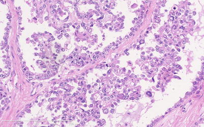
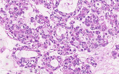
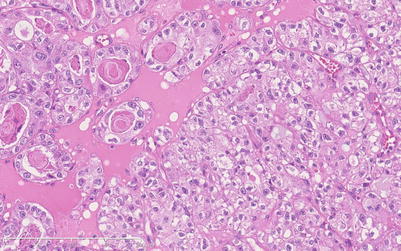
Fig. 4.1
Solid field within an ovarian CCC consisting exclusively of oxyphil cells
Fig. 4.2
Another tumor with another broad field of oxyphil cells
Fig. 4.3
At higher magnification, the amphophilic cytoplasm of these oxyphil cells is easily noted
Fig. 4.4
Predominantly micropapillary and tubulocystic CCC with a field consisting exclusively of oxyphil differentiation
Fig. 4.5
Another pure oxyphil cells field in a CCC
Fig. 4.6
Higher-power magnification of amphophilic, granular-appearing cytoplasm within oxyphil cells. Also note nuclear atypia
Fig. 4.7
Solid field of oxyphil cells infiltrated by some lymphocytes and plasma cells
Fig. 4.8
Tumor field in which many individual cells show transitions from clear to oxyphilic cytoplasm
Fig. 4.9
Field in which oxyphil cells at the center are surrounded by clear cells on both sides, with some individual cells in transition
Fig. 4.10
Higher-power view of a tumor field in which individual cells show both oxyphilic and clear cell features
Fig. 4.11
Another tumor containing cells intermediate between clear cells and oxyphil cells
Fig. 4.12
More “hybrid” oxyphil/clear cells in a CCC
Fig. 4.13
Another tumor with cells showing both clear and oxyphilic features
Signet ring cells (Figs. 4.14, 4.15, 4.16, 4.17, and 4.18) are frequently present in small numbers but in our material were prominent in less than 10 % of cases. The intracytoplasmic inclusions may appear clear (Fig. 4.16), mucoid (Fig. 4.17), eosinophilic (Figs. 4.14 and 4.15), or basophilic (Fig. 4.18) in hematoxylin- and eosin (H&E)-stained preparations and usually compress the nucleus to one side of the cell.
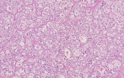
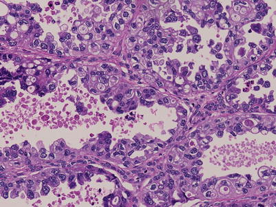
Fig. 4.14
Numerous signet ring cells (SRCs) containing eosinophilic globular inclusions
Fig. 4.15


Solid CCC field composed predominantly of SRCs with eosinophilic inclusions
Stay updated, free articles. Join our Telegram channel
Full access? Get Clinical Tree
