http://evolve.elsevier.com/Edmunds/NP/

 Top 100 drug;
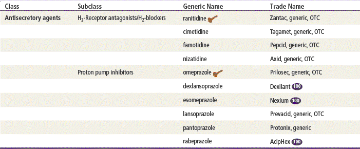
  key drug. Ranitidine is the most commonly used H2-receptor antagonist. Omeprazole is considered the key PPI because it is was the first to become available OTC and generic.
H2-blockers (H2-receptor antagonists) and PPIs block acid secretion in the stomach but do so via different mechanisms of action. In general, all H2-blockers, except cimetidine, are considered equally effective and have similar side effect profiles. The indications and off-label uses for each agent are listed in Table 27-1. These distinctions are not always observed in practice.
PPIs generally are considered to be more powerful in terms of action than H2-blockers because they decrease stomach acidity to a greater extent. Six PPIs are available, and little difference between them has been noted, except for duration of action.
Therapeutic Overview
Several protective factors work together to protect the stomach mucosa from injury. The gastric mucosal barrier resists backward diffusion of hydrogen ions and thus has the ability to contain a high concentration of hydrochloric acid within the gastric lumen, unless an injurious agent breaks the barrier.
Endogenous prostaglandins are thought to provide cytoprotection against injurious agents and are synthesized abundantly in the mucosa of the stomach and duodenum. They are known to stimulate secretion of both mucus and bicarbonate and to maintain mucosal blood flow.
Mucus also mediates mucosal protection. It is secreted by surface epithelial cells and forms a gel that covers the mucosal surface and physically protects the mucosa from abrasion. It also resists the passage of large molecules such as pepsin. Bicarbonate is produced in small amounts by surface epithelial cells and diffuses up from the mucosa to create a thin layer of alkalinity between the mucus and the epithelial surface. Other protective factors include mucosal blood flow, epithelial renewal, and epidermal growth factor that is secreted in saliva and by the duodenal mucosa.
The mucosal surface of the stomach is divided according to type of gland—namely, the oxyntic (parietal) gland area—which secretes acid, and the pyloric gland area, which does not. The most important cell types in these glandular areas are mucous and peptic cells, which are found in both glandular areas; oxyntic cells, which occur only in the oxyntic gland area; and endocrine cells, which are scattered throughout both glandular areas. The oxyntic gland area occupies most of the mucosal surface of the stomach. The oxyntic cells secrete HCl through an energy-dependent active transport mechanism, along with intrinsic factor, a protein required for the absorption of vitamin B12.
The stomach has three naturally occurring secretagogues that stimulate acid secretion. These include acetylcholine (a neurotransmitter), gastrin (a hormone), and histamine (a paracrine substance). Receptor activation by these secretagogues initiates biochemical steps that lead to active transport of hydrogen by the oxyntic cell. Histamine, released by mast cells, binds to the H2-receptor that activates intracellular stimulatory protein G and eventually activates membrane-bound adenosine triphosphatase (ATPase). This proton pump provides the energy to extrude hydrogen from the oxyntic cell in exchange for potassium. In this manner, histamine initiates a train of intracellular events that stimulate the continuous secretion of acid.
The rate of secretion varies greatly, depending on the time of day or night and the proximity to the time that a meal was eaten. When the stomach is at rest, the rate of secretion is low. This basal rate contrasts greatly with the high rate of secretion found at mealtime. Basal secretion in the human stomach exhibits a circadian rhythm, characterized by a maximal rate in the evening and a minimal rate in the morning.
The high rates of acid secretion surrounding mealtimes are reduced to the basal rate of secretion through inhibitory processes that begin to occur during a meal. Abatement of hunger suppresses the cephalic phase of gastric secretion. Secretion of HCl by oxyntic cells causes antral mucosal surface pH to fall to below 3.0, prompting the release of the paracrine substance somatostatin. Somatostatin decreases gastrin release from G-cells and directly inhibits oxyntic cell secretion of acid.
Pathophysiology
Drugs that decrease acid production, or antisecretory agents, are used to prevent the autodigestion of the upper GI tract by the acid–pepsin complex. Drugs or foods that increase acid production may provoke autodigestion. This is often the pathogenesis of peptic ulcer disease (PUD). These agents not only cause injury themselves but also augment the injury initiated by other agents.
The mucosa of the upper GI tract is susceptible to injury from a variety of conditions and agents. Endogenous agents include acid, pepsin, bile acids, and other small intestine contents. Exogenous agents include ethanol, aspirin, and NSAIDs.
Acid is essential for the occurrence of peptic injury. A pH of 1 to 2 maximizes the activity of pepsin. In addition, mucosal injury from aspirin, other NSAIDs, and bile acids is augmented in the presence of acid. On the other hand, ethanol causes mucosal injury with or without acid. Corticosteroids, smoking, and physiologic and psychologic stress predispose some people to mucosal injury via mechanisms that are not completely understood. When negative feedback is overwhelmed or is ineffective, erosion (<5-mm lesion) or an ulcer (>5 mm in diameter) may result. Ulcers represent loss of the enteric surface epithelium that extends deeply enough to reach or penetrate the muscularis mucosae. Because pepsin is active only at a pH of 1 to 4, neutralization of acid or inhibition of acid production eliminates the harmful effects of pepsin.
Disease Process
Peptic ulcer is defined as a break in the gastric or duodenal mucosa that extends through the muscularis mucosae. It arises when the normal mucosal defensive factors are impaired or overwhelmed. Duodenal ulcers are more common between the ages of 30 and 55 years and in males; gastric ulcers are more common between the ages of 55 and 70 years. Ulcers are five times more common in the duodenum than in the stomach. Duodenal ulcers are almost never malignant, whereas 3% to 5% of gastric ulcers are malignant.
Three important causes of PUD have been identified: NSAIDs, H. pylori, and acid hypersecretory states such as Zollinger-Ellison syndrome. The provider should confirm the presence of H. pylori and NSAID use before determining what therapy is warranted.
Mechanism of Action
Medications that inhibit acid secretion can act in several ways at several locations on or in the oxyntic cell. H2-receptor antagonist drugs can bind to the H2-receptor, thereby displacing histamine from receptor binding sites and preventing stimulation of the oxyntic cell by the secretagogue. The four H2-receptor antagonists have similar chemical structures. They reversibly inhibit basal or histamine-, pentagastrin-, or meal-stimulated acid secretion in a linear, dose-dependent manner by interfering with histamine at the H2-receptors on the gastric parietal cells. As much as 90% inhibition of vagal and gastrin-stimulated acid secretion occurs with these agents, reflecting the importance of histamine in the mediation of cholinergic and gastrin-stimulated acid secretion. Near-complete inhibition of nocturnal acid secretion may be achieved as well.
PPIs irreversibly inhibit the acid secretory pump embedded within the parietal cell membrane by altering the activity of hydrogen potassium ATPase (H+/K+ ATPase). This enzyme affects the secretory pump in the gastric parietal cell by inhibiting hydrogen ion transport into the gastric lumen and by decreasing stimulated acid secretion. It decreases the volume of gastric fluid. An increase in serum gastrin concentrations is observed. Because PPIs act on the basolateral membrane of the parietal cell, they do not affect gastric emptying, basal or stimulated pepsin output, or secretion of intrinsic factor. This type of drug does not seem to affect the ATPase of other organ systems.
Treatment Principles
Evidence-Based Recommendations
Cardinal Points of Treatment
Nonpharmacologic Treatment
Lifestyle modifications are integral to treatment for PUD. Recommendations no longer include bland or restrictive diets. Patients should eat balanced meals at regular times and should avoid foods that exacerbate symptoms. High fiber is encouraged. Caffeine causes increased acid secretion and should be avoided. Alcohol can aggravate an ulcer but usually does not appear to be harmful when taken in moderation. Smoking should be strongly discouraged.
Pharmacologic Treatment
Goals include relief of symptoms and healing of the ulcer. Treatment consists of a course of an H2-blocker or a PPI. In general, PPIs are used first line because they are more potent and require a shorter course of treatment. H2-blockers are less expensive and usually are also effective. If the patient does not start to see improvement within a few days, increase the dose of medication or change from an H2-blocker to a PPI.
H2-blockers (e.g., ranitidine, famotidine, cimetidine) are associated with 70% to 80% healing of duodenal ulcers after 4 weeks, and with 87% to 94% healing after 8 weeks. PPIs should be given for 4 weeks for a duodenal ulcer and for 8 weeks for a gastric ulcer and are associated with 80% to 100% healing. See Table 27-2 for recommendations on therapy details.
TABLE 27-2
| Type of Medication | Indication | Length of Time |
| H2-blockers | Duodenal ulcer | 8 wk |
| Eradication of H. pylori | Short term | |
| Erosive esophagitis | Long term | |
| Gastric ulcer | 12 wk | |
| GERD | 12 wk | |
| Heartburn | Short term | |
| Hypersecretory conditions | Long term | |
| Prevention of gastric NSAID damage | Long term | |
| Upper GI bleeding | Short term | |
| Proton pump inhibitors | Active duodenal ulcer | 4-8 wk |
| GERD | 4-8 wk | |
| Hypersecretory conditions | Long term |
Stay updated, free articles. Join our Telegram channel

Full access? Get Clinical Tree


