24 Hip and Thigh Fractures
Anatomy of the Hip and Thigh
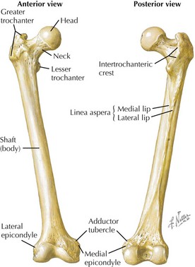
Femur
• Parts and landmarks: head; fovea (for round ligament); neck; greater trochanter; lesser trochanter; intertrochanteric line, crest, and fossa; pectineal line; gluteal tuberosity; linea aspera; shaft (body); popliteal surface; adductor tubercle; medial epicondyle; lateral epicondyle; medial condyle; lateral condyle; intercondylar fossa; patellar surface
Coxal (Hip) Bones
• Ilium, ischium, and pubis are fused in adults. (See Chapter 17, Pelvic Fractures, for more bone information.)


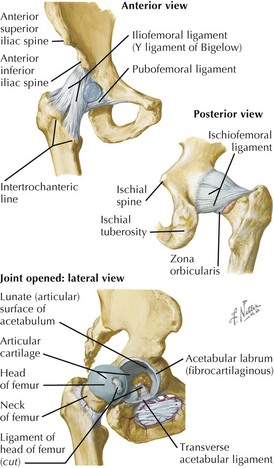
Hip Joint


• (Collateral) ligaments: spiraling thickenings of fibrous joint capsule, passing from acetabular rim to intertrochanteric line or trochanters
 Iliofemoral (Bigelow): anterior-superior, Y-shaped, very strong, prevents hyperextension by screwing femoral head tightly into acetabulum
Iliofemoral (Bigelow): anterior-superior, Y-shaped, very strong, prevents hyperextension by screwing femoral head tightly into acetabulum


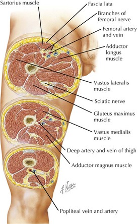
Compartments of the Thigh


• Fascia lata: investing deep fascia of thigh
 Attaches proximally to inguinal ligament, pubic rim, Scarpa’s fascia, iliac crest, sacrum, coccyx, sacrotuberous ligament, ischial tuberosity
Attaches proximally to inguinal ligament, pubic rim, Scarpa’s fascia, iliac crest, sacrum, coccyx, sacrotuberous ligament, ischial tuberosity


