Herpes Simplex Virus Esophagitis
Laura Webb Lamps, MD
Key Facts
Etiology/Pathogenesis
Esophagus most common site of infection
HSV almost exclusively infects squamous epithelium
Infection most often seen in immunocompromised patients
Macroscopic Features
Earliest lesion is shallow vesicles in mid to distal esophagus
Vesicles slough to become ulcers
Microscopic Pathology
Ulceration
Exudate with sloughed epithelial cells
Characteristic nuclear inclusions may be Cowdry type A or B
Diagnostic Checklist
Viral culture, immunohistochemistry, and molecular testing very helpful in resolving differential diagnosis
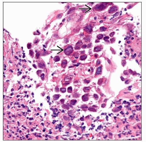 This case of herpes simplex virus (HSV) esophagitis features a dense neutrophilic exudate with admixed sloughed degenerating epithelial cells containing ground-glass HSV viral inclusions  . . |
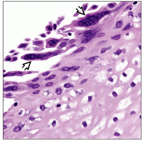 The squamous epithelial cells in herpetic esophagitis may contain multiple herpes simplex virus (HSV) inclusions  (polykaryons) within the surface epithelium. (polykaryons) within the surface epithelium. |
TERMINOLOGY
Abbreviations
Herpes simplex virus (HSV)
ETIOLOGY/PATHOGENESIS
HSV Esophagitis
Esophagus is most common site of infection in pediatric patients
HSV almost exclusively infects squamous epithelium, so areas of the alimentary tract lined by glandular epithelium are typically not affected
Most often seen in immunocompromised patients
Believed to represent reactivation of latent infection acquired earlier in life
Common pathogen in children with HIV
Primary infection in healthy patients may cause a self-limited esophagitis
HSV1 and HSV2 infection produce identical pathologic features


