11 Hernias
Anatomy of the Abdominal Wall
Abdominal Wall Layers
• Following are layers from the surface in.
 Superficial fascia with a variable amount of subdermal fat: Camper’s fascia, overlying membranous Scarpa’s fascia (subumbilical level)
 Muscle bellies and aponeuroses of the rectus abdominis, external and internal obliques, and transversus abdominis muscles
 Superficial fascia with a variable amount of subdermal fat: Camper’s fascia, overlying membranous Scarpa’s fascia (subumbilical level)
 Muscle bellies and aponeuroses of the rectus abdominis, external and internal obliques, and transversus abdominis muscles
External Oblique (EO) Muscle
• On each side, the lower border of its aponeurosis attaches to anterior superior iliac spine and pubic tubercle to form the inguinal ligament.
• Distally, a portion of EO aponeurosis fibers arch posteriorly to insert on the superior pubic ramus, forming the lacunar ligament (of Gimbernat).
• Most lateral of these deep (lacunar) fibers continue to run along the pectin of pubis as the pectineal ligament (of Cooper).
• Some of the most distal fibers arch upward, avoid the pubic tubercle, and merge with the opposite side’s fibers as the reflected inguinal ligament.
• Most muscle and aponeurotic fibers run superolateral to inferomedial (“hands in pockets” orientation).
• Superficial (external) inguinal ring: division in the most inferior aponeurosis; spermatic cord or round ligament passes through
Internal Oblique (IO) Muscle
• Fibers run deep, approximately perpendicular to the external oblique layer, from the deep lumbar aponeurosis, curving anteriorly then medially.
• Medial IO aponeurosis layer splits to pass around the rectus, as the middle layer of the rectus sheath, above the semicircular lines (of Douglas).
• On each side, the inferior triangle of the IO aponeurosis fuses with the transversus aponeurosis to form the conjoined (conjoint) tendon.
Transversus Abdominis (TA) Muscle
• Fibers run deep to the internal oblique layer, mostly posteriorly, becoming largely aponeurotic laterally in the deep back.
• Medial aponeurotic fibers pass posterior to the rectus, as the posterior layer of the rectus sheath, above the semicircular lines (of Douglas).
• On each side, the inferior triangle of the TA aponeurosis fuses with the internal oblique aponeurosis to form the conjoined (conjoint) tendon.
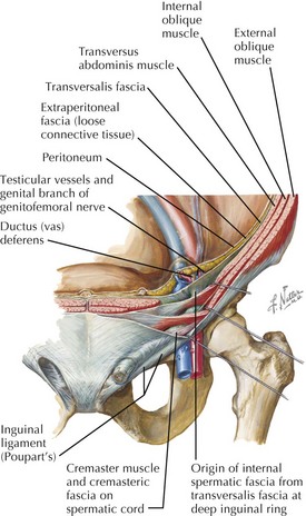
• Deep (internal) inguinal ring: gap in the transversus abdominis, lateral to the inferior epigastric arteries
Rectus Abdominis Muscle
• Parallel segments of muscle with vertically running fibers; segments joined end-to-end by tendinous insertions (inscriptions)
• External oblique aponeurosis is always the most superficial (anterior) component of the rectus sheath.
• Internal oblique aponeurosis splits to run in front of and behind rectus in the sheath above the semilunar lines (somewhat above umbilicus).
• External and internal oblique and transversus aponeuroses components of rectus sheath pass anterior to the rectus below the semicircular lines (below the umbilicus).
Linea Alba
• Midline, tendinous junction between right and left portions of the rectus sheath and the underlying midline tendons of the rectus muscle segments
Transversalis Fascia
• Tough fascial layer just deep to the transversus muscle and aponeurosis, rectus sheath, and rectus abdominis anteriorly
• Male transversalis fascia outpockets through the deep (internal) inguinal ring, a gap in the transversus abdominis, lateral to the inferior epigastric arteries.
• Internal spermatic fascia: transversalis fascia layer surrounding the layers of the tunica vaginalis around the descended testis, its duct, and vessels
Stay updated, free articles. Join our Telegram channel

Full access? Get Clinical Tree


