CHAPTER 31 Hemostasis
Principles of investigation
Introduction
The hemostatic system, a complex defense against bleeding, is critical to survival. Its integrity is compromised by inherited or acquired failure of its individual components, or by deregulation of the entire system provoked by organ failure, the inflammatory response, or exposure to cancer cell surfaces. Hemostasis also acts (in the wrong place, at the wrong time) as thrombosis. Bleeding is always a threat, while thrombosis increases due to age-related changes in coagulation factors and blood vessels to become the dominant hemostatic risk in later life.
The extreme complexity of hemostasis revealed by scientific scrutiny induces a degree of alienation in many clinicians practicing at the bedside and in the operating theate. The hematologist must be their translator of basic knowledge into clinically useful advice, and guide to the increasing menu of potent drugs and biological agents available for the therapy of bleeding and thrombosis.
To do this work a reliable toolkit of investigational methods is essential. These include a focused approach to the patient’s personal and familial medical history, a set of rapid laboratory tests to indicate the presence and general nature of any hemostatic malfunction, and the ability to extend this inquiry to measurement of specific proteins and analysis of DNA if required. The principle underlying these ‘nested’ methods of investigation is common to all disciplines in clinical pathology: provide data that increases (or decreases) the likelihood that a particular pathologic state – a diagnosis – is present and needs specific therapy or other intervention.
Hemostatic tests retain unique features and problems in interpretation. Even coagulation screening tests (the only commonly requested tests that require explicit coreporting of control experiments) are complex bioassays in miniature. An abnormal value can have diametrically opposed meanings for patient care depending on the clinical context. Expressing clinical pretest probability in an intelligible way and using test results to modify this probability is the best way of avoiding potential confusion and error.1
The application of meta-analysis of randomized studies (‘evidence-based medicine’) to diagnostic laboratory testing has been limited,2 and hemostatic testing is no exception. It is therefore not possible yet to claim evidence-based validation, in its strict sense, for many of the principles discussed below. However, the writings of many expert clinician–scientists over the years are the best guide we have to these principles, and should certainly form a starting-point for further analyses.
Physiology of hemostasis applied to diagnosis
The clinical approach to the patient who may have a hemostatic disorder is informed by knowledge of the physiology of hemostasis. Hemostatic reactions operate in a clock-like sequence, the first two phases being termed ‘primary’ and ‘secondary’ hemostasis.
A careful clinical history and examination (see below) can tentatively locate the potential defect in one of these phases, guiding the selection of initial investigations. The pretest probability of a defect involving primary hemostasis rises if abnormal bleeding follows a ‘mucosal’ pattern (see below), while a history of muscle or joint bleeding increases the likelihood of a coagulation deficiency. Disorders of the regulatory protein C pathway tend to manifest as venous thromboembolism. Abnormalities of the final phase of hemostasis, fibrinolysis, tend to contribute to bleeding in specific clinical settings, for example disseminated intravascular coagulation (DIC) and hepatic failure.
To assist this diagnostic thinking, it helps to keep in mind a simplified map of the hemostatic system, whatever knowledge of its complexity one possesses (or not, as the case may be). These simple maps are caricatures: readers are referred to fuller versions3,4 and to other chapters in this volume.
Primary hemostasis: formation of the platelet plug5 (Fig. 31.1)
Platelet adhesion
Vessel wall damage provokes a variety of signals from damaged or activated vascular endothelial cells and/or exposure of the underlying subendothelial matrix. Platelets first adhere to the signaling site via adhesive ligands. The dominant interaction is between platelet membrane receptor glycoprotein Ib-IX and the giant polymer von Willebrand factor (vWF), particularly its most adhesive high molecular weight forms.
Platelet aggregation
Adherent platelets flatten and activate membrane fibrinogen receptors (glycoprotein IIb-IIIa) that bind plasma fibrinogen (Fb). The resulting syncytial platelet aggregate provides a reactive surface composed of platelet membranes. This activated membrane flips inside out, exposing the negatively charged phospholipid, phosphatidylserine (PS). By binding the tenase and prothrombinase complexes, PS allows coagulation reactions to proceed. This altered (‘activated’) platelet membrane is sometimes termed platelet factor 3.
Secondary hemostasis: generation of fibrin clot by the coagulation pathway (Fig. 31.2)
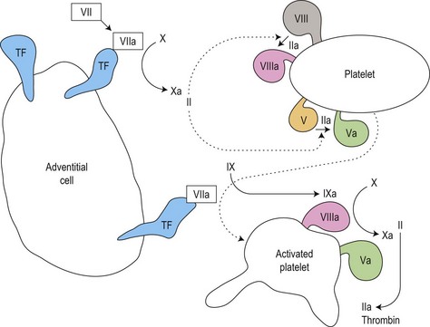
Fig. 31.2 Thrombin generation in vivo: coagulation factor interactions mediated by cell surfaces (schematic).
Clot initiation: the tissue factor/factor VIIa complex
The receptor tissue factor (TF), exposed on adventitial cells, activated endothelial cells and leukocytes in the damage zone, then binds and activates factor VII. TF/VIIa complexes bind and activate factor X. Resulting FXa moves to the platelet surface.
This is a regulatory ‘decision point’. If this burst of FXa cleaves sufficient thrombin from its precursor (prothrombin), thrombin-mediated activation of the co-factors factor VIII and factor V, and the enzyme factor IX – together with recruitment of more thrombin-activated platelets – assemble a ‘critical mass’ that allows coagulation to proceed. If thrombin generation falls short, tissue factor pathway inhibitor (TFPI) suppresses the TF/VIIa/Xa complex and coagulation is stalled.
Clot amplification: the ‘tenase’ complex
If the ‘decision’ is positive, sufficient FVIIIa and FIXa are formed to make the intrinsic tenase complex, in which FVIIIa acts as a rate-enhancing co-factor in the cleavage of FX to FXa by FIXa, providing a sustained source of FXa. The location of this FXa on the platelet surface enables it to move to the nascent prothrombinase complex.
Clot propagation: the prothrombinase complex
The surge of FXa forms prothrombinase complexes with FVa on platelet surfaces, speeding thrombin generation from prothrombin. Thrombin cleaves fibrinogen to form a durable fibrin clot, and binds to it, promoting further clot growth. This is a secure barrier against bleeding.
Clot regulation and removal: the protein C and fibrinolytic pathways
Two further systems regulate and eventually remove the clot (in the context of tissue repair and neoangiogenesis) (also see Chapter 28):
Clot regulation: the protein C system6 and antithrombin
Thrombin activity is restricted to the platelet surface and the fibrin matrix of the clot by a conformation-dependent inhibitor, antithrombin (AT)7 that inactivates fluid-phase thrombin. To work efficiently, AT must bind to heparin-like proteoglycans on healthy endothelial cells.
Fibrinolysis: the plasmin system8
Clots contain the seeds of their own destruction in the form of a clot-bound protein, plasminogen. This is cleaved by tissue plasminogen activator (tPA) secreted by healthy vascular endothelial cells, or urokinase on the surface of macrophages, to the fibrinolytic enzyme plasmin. Plasmin cleaves fibrin into fibrin degradation products, notably the D-dimer fragment specific to cleavage of cross-linked fibrin.9 This removes the clot, and activates matrix metalloproteinases that initiate remodeling of vessels (angiogenesis).
The clinical approach to the patient with a possible hemostatic disorder
The question of a possible hemostatic disorder occurs in two main settings. An individual is referred because they have presented with, or self-reported, clinical phenomena suggesting excess bleeding. Investigation can proceed in a structured elective style. In the second case, excess bleeding occurs acutely in a patient undergoing treatment in the hospital, emergency department or surgical theater. The tempo, urgency and completeness of the diagnostic work-up (before recourse to therapeutic action) are then different, but the principles are shared.
Experts writing about the investigation of possible bleeding disorders unanimously stress the importance of a carefully taken history.10–12 They also recommend specific questions, answers to which alter the pretest probability of a bleeding disorder. The discussion below draws on this consensus. Similarly, key findings on clinical examination may aid the diagnostic process, although they are less frequent than narrative clues.
It must be conceded that these narrative and clinical signs have not been formally tested, either singly or in clusters, for their relative value in predicting the presence of hemostatic disorders. Such testing has refined and simplified the use of clinical clues in other contexts,13 and may be of future benefit in hemostasis. Until such clarification becomes available, the shared insight of experienced clinicians is our best guide.
History
The role of the history-taker is to determine if the patient’s account is consistent with excessive bleeding. After initial open questioning related to the presenting complaint, a systematic inquiry is made with the help of key questions intended to elicit quantitative information about the bleeding in question. People (including doctors) tend to overestimate, by eye, volumes of blood lost from the body, so it is more informative to focus questioning on the duration of a bleeding episode and what had to be done about it.
Key questions
Surgical challenges
Dental surgery
Surgical trauma to the incompressible tooth socket sitting in the fibrinolytic milieu of the oral cavity is a stiff challenge to the hemostatic system. Useful questions about the effect of extractions focus on the duration of bleeding and the actions compelled by it. Compare the two accounts in Box 31.1. The second account gives a much clearer indication of excessive blood loss. Most people (in the UK, at least) are disinclined to make an early return to the dentist without a pressing reason.
Other types of surgery
Questions about blood loss after circumcision and tonsillectomy are traditional, but the timing and selectivity of the former, and decreasing popularity of the latter, mean that only a small minority of individuals (or their parents) will give a useful response. As in the case of dental extraction, questions should focus on duration of bleeding and subsequent medical actions.
Many individuals are referred for investigation of a possible bleeding disorder as a result of excess blood loss after major surgery, although the commonest cause of this is purely ‘surgical’ – a transected blood vessel evading the hemostat. Large and/or late wound hematomata or generalized ‘oozing’ from a tissue surface or organ bed are more likely to indicate a hemostatic disorder. The patient’s own recall of these events is likely to be hazy, and documentation of the amount, duration and clinical reaction to peri- and postoperative bleeding should be sought in the patient’s medical records.
Epistaxis
Nosebleeds are a universal experience in childhood, so the usefulness of enquiring about them (nearly everyone will recall some) depends on the questions asked. In bleeding disorders, epistaxes tend to run ‘like a tap’ rather than to drip; require a bowl to catch the blood rather than a tissue; and resist arrest (or reroute via the mouth) on pinching the nares. Frequently recurring epistaxes that provoke multiple nasal cauterizations also increase the possibility of a bleeding disorder.
Gastrointestinal or urogenital bleeding
Rectal bleeding compels a search for colorectal disease even if a systemic bleeding disorder is present. Coumarin-induced rectal bleeding has led to the early detection and cure of cancers. Similar action must follow hematemesis, hematuria or vaginal bleeding. Occasional prolonged episodes of spontaneous hematuria occur in hemophilia, sometimes in mildly affected individuals.
Menstruation
As with other perceptions of bleeding symptoms by both sexes, women accustomed only to their own menstrual loss may not regard it as abnormally heavy. Bleeding for >7 days per month, bleeding that regularly ‘breaks through’ sanitary protection, the need to wear both tampons and pads (or double pads), and the need to protect the bed with a towel, or to cancel social engagements due to bleeding, are reliable indications of menorrhagia. Questioning should be sensitive and preceded by an explanation of its relevance.
Bruising
A sizeable minority of the population will answer ‘yes’ to ‘do you bruise easily?’ and many older people bruise the sun-thinned skin of their hands and forearms, so this question is not helpful. A semi-quantitative approach is useful: the bruises can be compared to some common object (in the UK the 50 pence coin, about 2 cm in diameter). Having frequent bruises larger than this is significant. Most normal (’simple’) bruises occur on the outer surfaces of the upper arms and thighs, ‘bumpers’ in contact with the environment: bruises on the trunk, neck or face, or on the inner aspects of limbs are more significant, as are palpable bruises (hematomata). Solar or simple bruises are rarely pathologic.
A patient complaining of easy bruising who cannot show a single bruise at the time of the consultation, or one who agrees that there are fewer days with bruises than days without,11 is less likely to have a bleeding disorder. Thrombocytopenic purpura crop around the ankles, where venous pressure is highest, and are more likely to be perceived as ‘a rash’ than as bruises.
’Third space’ bleeds
In the hemophilias (inherited and acquired), over-anticoagulation with heparins or coumarins, and other systemic bleeding disorders, the presenting complaint may be hemorrhage into joints, muscles, or other deep tissue compartments. These events may not be perceived as bleeds by the patient or even by the attending clinical team, since they present with pain, swelling, nerve entrapment or other space-occupying features rather than with evident blood loss. By mimicking tumors, or presenting as acute monoarthritis, they may provoke biopsy or drainage attempts with potentially catastrophic results. In the context of anticoagulant therapy, failure to recognize such bleeds, and consequent ‘pushing on’ with heparin or warfarin, is equally dangerous.
The first-line clinicians called upon to deal with these events are rarely experienced in their recognition, so the best protection lies in local in-service education and guidelines, together with constant availability of hematologic advice and the freedom to access it.
Summation and duration of bleeding episodes
All types of blood loss should be summated. A patient with a credible history of significant bruising and epistaxis is more likely to have a bleeding disorder than one with bruising alone. Bleeding symptoms that go back to childhood or adolescence are likely to be inherited, and prompt a family history, while if recently developed they point to an acquired cause and a general enquiry for systemic disease.
Pattern of bleeding
A ‘mucosal’ pattern of bleeding episodes (epistaxis, menorrhagia, bleeding after dental surgery) may guide the initial investigation towards platelet and vWF analysis since it suggests a problem with primary hemostasis. Presentation with hemarthrosis or other third-space bleeds is classical in hemophilia. However, this is hardly a clear distinction, since hemophilia also causes mucosal hemorrhage and dental disasters: stating that menorrhagia is more likely in primary bleeding disorders than in hemophilia is tautologic. In general, it is necessary to perform at least screening tests (see below) of both primary hemostasis and coagulation in people who bleed too much.
Drug history
A full list of all prescribed or over-the-counter medication (including herbal and other complementary medicines) taken by the individual should be compiled. Aspirin remains the most prevalent agent causing bleeding symptoms and abnormal platelet function test results: in addition to being prescribed widely for its antithrombotic effect, it is a component of many preparations on sale to the public. Some of these preparations have names that advertize the presence of aspirin (e.g. Aspro®) while others (e.g. Nurse Sykes’ Powders®) do not: a full list of such products is given in the British National Formulary.14 Other non-steroidal anti-inflammatory agents share the aspirin effect. Antibiotics, major tranquillizers and antidepressive agents may all be associated with bleeding via antiplatelet function effects. Platelet function testing should be performed first with the patient taking the drug, then 2 weeks after stopping, in order to demonstrate its effect.
Family history
A reliable family history entails documenting a pedigree chart including all known family members with their names and dates. The key questions illustrated above are asked about each member in turn, seeking confirmation of any said to have a bleeding tendency. Any described as having hemophilia should, if possible, be traced to the Hemophilia Center carrying out their care: a relative famous for ‘hemophilia’ often turns out to have no evidence of the disorder at all.
Taking a family history of this quality is time-consuming, may take more than one session, and suits the elective better than the emergency setting. It often extends the individual’s historical knowledge of their family beyond its limit. Furthermore, a negative family history excludes nothing, since many bleeding disorders, including severe hemophilia, occur sporadically.
Clinical examination
Skin
The whole skin surface should be inspected for purpura and bruising, documenting the distribution, size and age of lesions and correlating them with the clinical history. Palpation of bruises will detect hematomata, while palpable purpura suggests vasculitis. Close attention should be paid to the ankles, where venous and capillary pressure is highest: petechiae first appear here in thrombocytopenia, and signs of venous or arterial insufficiency may be evident. Large bruises (ecchymoses) typical of hemophilia or anticoagulant overdose may be found tracking into dependent parts of the body such as the scrotum.
The surface of lesions should be inspected. Edema may indicate the urticarial component of anaphylactoid purpura. Lesions of hereditary hemorrhagic telangiectasia may be seen in finger pulps and ear lobes, spreading over the face in later life. Bruises with abrasions or thermal trauma, that follow the outline of a blunt object, or are associated with other signs of abuse or self-harm may indicate non-accidental injury or factitious bruising.
Scars should be examined. Keloid formation might rule out a skin bleeding time. In Ehlers–Danlos syndrome they pucker like tissue paper on sideways compression, and may show central breakdown with fresh exudation. Poor scar quality may also be seen in hypo- or afibrinogenemia.
Non-hemorrhagic lesions mistaken for signs of bleeding include cherry-red Campbell de Morgan spots, stretch marks, livedo reticularis and Majocchi’s purpura or other ‘dermatological’ purpuras.
Mucosae
The oral cavity should be inspected for the petechiae or ‘blood blisters’ of ‘wet’ thrombocytopenia. Gingival bleeding is usually associated with gingivitis. Oral hemorrhage in the hemophilias occurs at sites of minor trauma or dental surgery, and may consist of a small but persistent bleeding point, a friable oozing clot, or a tumor-like sublingual swelling.
Musculoskeletal system
Joints should be examined for warmth, effusion, synovitis, reduced range of movement and misalignment. Muscle groups should be examined for wasting and contractures. These signs of cumulative damage due to hemarthrosis and intramuscular hematomas are characteristic of the hemophilias, but are also seen in rare disorders such as type 3 von Willebrand disease (vWD), deficiency or severe recessive disorders such as homozygous factor XIII, factor VII or factor X deficiency. Intramedullary hemorrhage of the long bones is a feature of afibrinogenemia and α2-antiplasmin deficiency, both very rare: it mimics lytic bone disease.15
Nervous system
Evidence of nerve compression injuries may be evident combined with damage to the musculoskeletal system identified above. Retinoscopy should be performed in all patients with purpura, particularly involving the oral mucosa: retinal hemorrhages indicate active CNS bleeding and the need for urgent therapy.
Active bleeding
The postoperative or traumatized patient with excessive bleeding should be examined for the signs itemized above, but sites of blood loss should be directly observed if possible. External losses, including those via surgical drains, should be assessed: dilution with tissue exudate can exaggerate blood losses. Similar overestimation can occur in hematuria. If in doubt in either of these situations, a hemoglobin estimate on the drain fluid or urine can be helpful. Tracking hematomata should be sought. All intravascular access points, together with other skin incisions pre- or postdating the main episode of blood loss, should be inspected for evidence of bleeding or rebleeding after earlier closure. Fresh bleeding from such sites is a sign of DIC in its consumptive phase.
On defining the pretest probability of a bleeding disorder
Using information from the history and examination the clinician can work out a broad pretest probability (e.g. low, moderate or high) that the patient has a clinical bleeding disorder. The accuracy and precision of the history and examination described above in defining this pretest probability have not been tested by methods that have provided such information in other contexts.13 Such studies are feasible and desirable in bleeding disorders, but even in their absence, a rational estimate of pretest probability is a crucial step towards interpreting the results of laboratory testing. Without it, tests of hemostasis can be frankly misleading.
Screening tests of hemostasis: two warnings
Armed with an estimate of pretest probability, the next step is to perform screening tests of hemostasis to generate further data capable of increasing or decreasing it.
On venipuncture
This requires a blood sample, which should be taken by an expert venipuncturist – especially in the case of a child – from a peripheral vein with minimal venous stasis. On no account should the jugular, subclavian or femoral veins be approached if there is any possibility of a bleeding disorder. Many inexperienced clinicians seem drawn to perform a ‘femoral stab’ on patients covered in bruises: this can result in a massive compartment bleed in the femoral triangle. Multiple attempts to obtain samples from the antecubital fossa can likewise result in severe bleeds. An expert hand is vital.
On screening tests
These tests ‘screen’ hemostasis, not people – a source of considerable misunderstanding and futile testing. They do not meet the epidemiological standard of true screening tests because they are not sensitive or specific enough to screen a population for bleeding disorder. They only work in concert with the history and examination as described above.
The 250 ‘clotting screen’ requests typically made every day in a large teaching hospital represent educational failure. This futile attempt to screen the population entering hospital for surgery (or other intervention) for bleeding risk depends partly on misinterpretation of the ambiguous term ‘screen’. Even more misleading – and potentially wasteful – is the lazy application of the term ‘thrombophilia screen’ to detailed testing for inherited and acquired thrombophilia. When the term ‘screen’ is unavoidable, it is used below strictly to refer to tests performed as the result of a clinical history of bleeding or thrombosis.
Initial screening tests, usually applied whatever the pattern of abnormal bleeding, consist of a multiparameter blood count including the platelet count, and coagulation tests: a prothrombin time (PT), activated partial thromboplastin time (APTT), and sometimes a thrombin clotting time (TT).
If the pretest probability of a bleeding disorder is possible or probable, normal results in these initial tests should be followed by a skin bleeding time estimation or whole blood platelet function analysis. The need for further platelet function tests, specific assays of hemostatic proteins or genes, or further clinical tests for systemic disorders depends in part on the results of ‘global’ tests of hemostasis, but should also proceed if the full history is convincing, even if initial tests are normal. Below, tests of primary hemostasis and coagulation are grouped together for coherency, but they are also ranked into ‘screening’ and ‘diagnostic’ categories.
Laboratory investigation of hemostasis
Tests of primary hemostasis
Screening tests
The platelet count
Methods. In the current laboratory, platelet counting is performed on an anticoagulated venous blood sample as part of the multiparameter ‘full blood count’ generated by automated cytometers. Current systems count particles of platelet-like size (2–37 µm3) by electrical aperture impedence or laser light scattering. To censor ‘noise’ at the low end and red cells at the high end of this range, devices fit a lognormal distribution curve to this raw count or otherwise manipulate it to calculate the reported platelet count.
The validity of the platelet count accordingly depends on instrument standardization, calibration and quality control: details of these procedures can be found elsewhere.15 Because instruments count particles by size, blast cell fragments (in acute leukemia) or schistocytic red cells (in thrombotic thrombocytopenic purpura) may lead to overestimation, and large platelets (in immune thrombocytopenia or myelofibrosis) to underestimation, of the true platelet count.
Normal and abnormal platelet counts. The normal (‘Gaussian’) reference range for the concentration of platelets in venous blood (’the platelet count’) is 150–400 × 109/l. By definition, 5% of normal individuals have platelet counts outside this range. To regard and investigate asymptomatic individuals with isolated, stable, mild thrombocytopenia (100–150 × 109/l) as if they had a disease may be to confound ‘Gaussian’ and ‘diagnostic’ concepts of normality.1 However, evidence to justify abandoning this seemingly unproductive practice is lacking.
Correlating the platelet count with the clinical situation. The action taken in response to the finding of a low platelet count depends on the presence or risk of bleeding, since the two are not always correlated. In many patients with immune thrombocytopenia (ITP), clinical bleeding may be minor or absent even at very low counts (<10 × 109/l), and precipitant therapy may not be necessary. However, the presence of mucosal bleeding in ITP indicates early therapy.
Lesser degrees of thrombocytopenia (20–50 × 109/l) are dangerous when combined with reduced platelet function (e.g. antiplatelet agents, myelodysplasia, myelofibrosis); abnormal coagulation (e.g. DIC); leukemia (e.g. acute promyelocytic leukemia); cerebral vasculopathy in sickle cell anemia, or with severe anemia of any cause. In these situations, aggressive therapy including intensive platelet transfusion support is often needed.
When confronting a reduced platelet count, an apparently simple variable, potential laboratory error or artifact must be sought, and the platelet count must be placed firmly in the clinical context. These are core principles in all hemostatic testing.

Stay updated, free articles. Join our Telegram channel

Full access? Get Clinical Tree