Heme Metabolism

Chapter 27 Heme Metabolism


Heme is a tightly bound prosthetic group of hemoglobin, myoglobin, the cytochromes, and other proteins. It is bound to its apoproteins either noncovalently, as in hemoglobin and myoglobin, or by a covalent bond, as in cytochrome c. Heme consists of a porphyrin, known as protoporphyrin IX, with an iron chelate in its center. The porphyrin system consists of four pyrrole rings linked by methine (—CH=) bridges:


The conjugated (alternating) double bonds absorb visible light. Therefore the heme proteins are colored. Porphyrinogens are important intermediates in heme biosynthesis. Their pyrrole rings are connected by methylene (—CH2—) bridges, and they do not absorb light because the double bonds do not alternate with single bonds over the whole system:


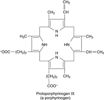
Porphyrinogens are prone to nonenzymatic oxidation to the corresponding porphyrins.


Although a small amount of dietary heme is absorbed in the small intestine, essentially all the heme in the human body is derived from endogenous synthesis. This chapter describes the pathways for the biosynthesis and degradation of heme and the diseases in which these pathways are disrupted.


Heme is synthesized from succinyl-coenzyme A and glycine


Heme biosynthesis starts with the formation of Δ-aminolevulinate from succinyl-coenzyme A (succinyl-CoA) and glycine, catalyzed by the heme-containing, vitamin B6–dependent enzyme δ-aminolevulinate (ALA) synthase. This is the committed step of the pathway shown in Figure 27.1.


image

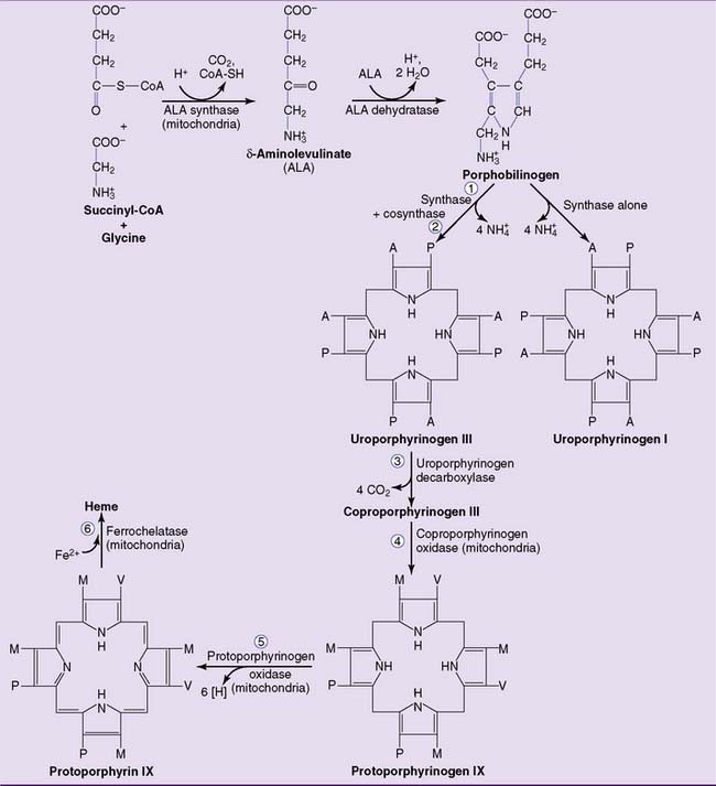
Figure 27.1 Pathway of heme biosynthesis. The numbered reactions refer to numbers listed in Table 27.1. A, Acetate (carboxymethyl) group; M, methyl group; P, propionate (carboxyethyl) group; V, vinyl group.


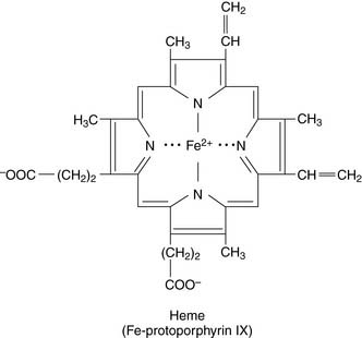
The second reaction, catalyzed by ALA dehydratase, forms the pyrrole ring of porphobilinogen. The third enzyme, uroporphyrinogen I synthase (also called porphobilinogen deaminase), links together four molecules of porphobilinogen. Left on its own, the synthase produces uroporphyrinogen I. In the cell, however, a second protein, uroporphyrinogen III cosynthase, channels the reaction into the formation of the isomer uroporphyrinogen III. All naturally occurring porphyrins, including heme, belong to the III series. Uroporphyrinogen III is processed to heme by the reactions shown in Figure 27.1.


The first reaction and the last three reactions of the pathway are mitochondrial. The others are cytoplasmic. ALA synthase is the regulated enzyme. ALA synthase has an unusually short biological half-life of 1 to 3 hours in the liver, and its synthesis is very effectively suppressed by heme. Only free, nonprotein-bound heme acts as a feedback inhibitor.


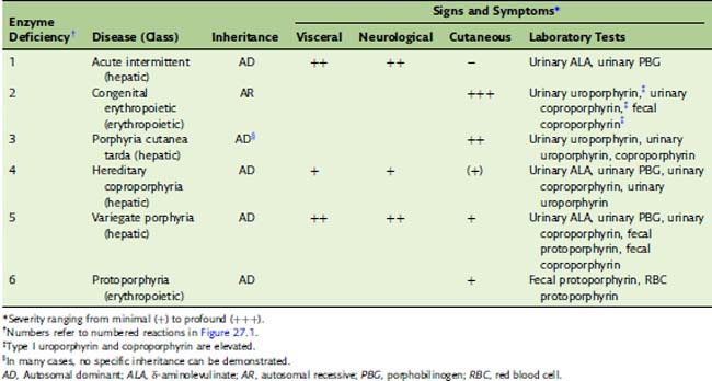
Porphyrias are caused by deficiencies of heme-synthesizing enzymes


The porphyrias (Table 27.1) are caused by a partial deficiency of one of the heme-synthesizing enzymes other than ALA synthase. Any one of the numbered enzymes shown in Figure 27.1 can be affected. A complete deficiency would be fatal, and the offending mutations are generally expressed as autosomal dominant traits; affected heterozygotes have 50% of the normal enzyme activity. Clinically, we can distinguish between hepatic porphyrias and erythropoietic porphyrias.


Clinical abnormalities in the porphyrias are caused not by heme deficiency but by the accumulation of metabolic intermediates. The substrate of the partly deficient enzyme accumulates in situations in which ALA synthase activity is high. Two types of toxic effect are especially common:


Two enzymes of heme synthesis, ALA dehydratase and ferrochelatase, are sensitive to inhibition by lead. This results in increased levels of urinary ALA and an increased concentration of protoporphyrin IX in erythrocytes. Some of the neurological impairments in lead poisoning are attributed to the accumulation of these metabolites in nervous tissue.


CLINICAL EXAMPLE 27.1: Acute Intermittent Porphyria


Acute intermittent porphyria (AIP) is caused by a dominantly inherited deficiency of uroporphyrinogen I synthase (porphobilinogen deaminase). The enzyme activity is reduced to 50% of normal in all tissues, but clinical manifestations are related to impaired heme synthesis in the liver because the activity of this enzyme, in relation to the other heme biosynthetic enzymes, is rather low in this organ.


Only 10% of individuals with the genetic trait ever show clinical manifestations. They experience episodes of abdominal pain, constipation, muscle weakness, and cardiovascular abnormalities. Agitation, seizures, or mental derangement may be present. These episodes last from a few days to several months and are attributed to the accumulation of aminolevulinic acid (ALA) and porphobilinogen in blood and cerebrospinal fluid. Although these metabolites originate in the liver, they cause most of the symptoms by acting on the central and peripheral nervous systems.


Acute attacks of AIP can be precipitated by barbiturates, phenytoin, griseofulvin, and other drugs that induce the synthesis of cytochrome P-450. When the P-450 proteins are induced by drugs, the small amount of free, unbound heme in the liver rapidly binds to the newly synthesized apoenzymes. This depletes the pool of free, unbound heme, and heme depletion derepresses ALA synthase. ALA and porphobilinogen accumulate when the activity of ALA synthase exceeds the activity of uroporphyrinogen I synthase.

Stay updated, free articles. Join our Telegram channel

Jun 18, 2016 | Posted by in BIOCHEMISTRY | Comments Off on Heme Metabolism

Full access? Get Clinical Tree

Get Clinical Tree app for offline access