FIGURE 17-1
(A) Cat-scratch lymphadenitis
(B) Fungal lymphadenitis
(C) HIV lymphadenitis
(D) Kikuchi lymphadenitis
(E) Tuberculous lymphadenitis
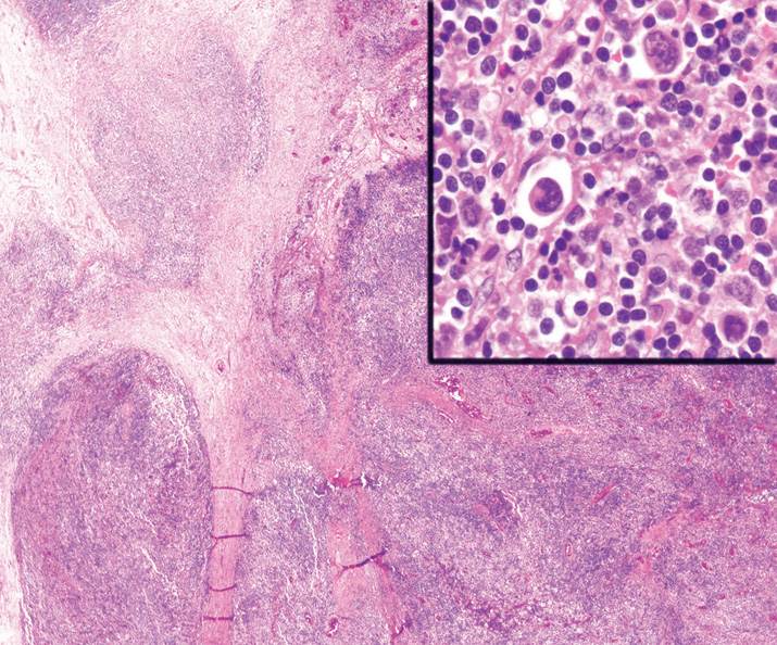
10. A 42-year-old man with a history of fever, diarrhea, and weight loss is found to have widespread lymphadenopathy. An image from his cervical lymph node excision is shown in Figure 17-2. Stains for acid-fast bacilli and fungal organisms were negative. A PAS-diastase stain was positive. These findings are diagnostic of
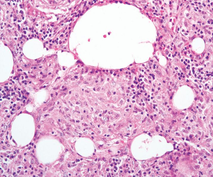
(A) Fungal lymphadenitis
(B) Hodgkin lymphoma
(C) Niemann-Pick disease
(D) Tuberculous lymphadenitis
(E) Whipple disease
11. All the following features are associated with toxoplasma lymphadenitis, except
(A) Aggregates of epithelioid histiocytes
(B) Aggregates of monocytoid cells surrounding blood vessels
(C) Follicular hyperplasia
(D) Necrosis
(E) Posterior cervical lymph node involvement
12. Which of the following carcinomas is least likely to metastasize to the supraclavicular lymph nodes?
(A) Colon
(B) Esophagus
(C) Lung
(D) Pancreas
(E) Stomach
13. All the following diseases cause matted lymphadenopathy, except
(A) Cat-scratch disease
(B) Cancer
(C) Lymphogranuloma venereum
(D) Measles
(E) Tuberculosis
14. All the following findings support a diagnosis of reactive lymphoid hyperplasia, except
(A) Distended germinal centers
(B) Enlarged, oddly shaped follicles
(C) Frequent mitoses
(D) Germinal centers are bcl-2 positive
(E) Peripheral rim of inactivated lymphocytes
15. Progressive transformation of germinal centers is associated with all the following findings, except
(A) Occurs in young adults
(B) Occasional Reed-Sternberg cells are present in the interfollicular space
(C) Presence of a large lymphoid nodule that is 3–5 times the diameter of an adjacent follicle
(D) Small lymphocytes infiltrate the germinal centers
(E) Usually presents as a single, enlarged lymph node
16. A 35-year-old man presents with a nodular lesion in the infraauricular region and an enlarged cervical lymph node. Lymph node examination shows diffuse eosinophilia with eosinophilic microabscesses and infiltration of the germinal centers. In addition, there is prominent vascular hyperplasia of post-capillary venules. Based on these findings, what is the best diagnosis?
(A) Angiolymphoid hyperplasia with eosinophilia
(B) Castleman’s disease
(C) Hodgkin lymphoma
(D) Kikuchi disease
(E) Kimura’s disease
17. A 14-year-old teenager with a history of massive lymphadenopathy and polyclonal hypergammaglobulinemia undergoes lymph node excision that shows marked sinusoidal dilatation with effacement of follicles. Emperipolesis is conspicuous. Which of following immunohistochemical stains is least likely to be expressed by the cells of interest?
(A) CD1a
(B) CD4
(C) CD30
(D) CD68
(E) S-100
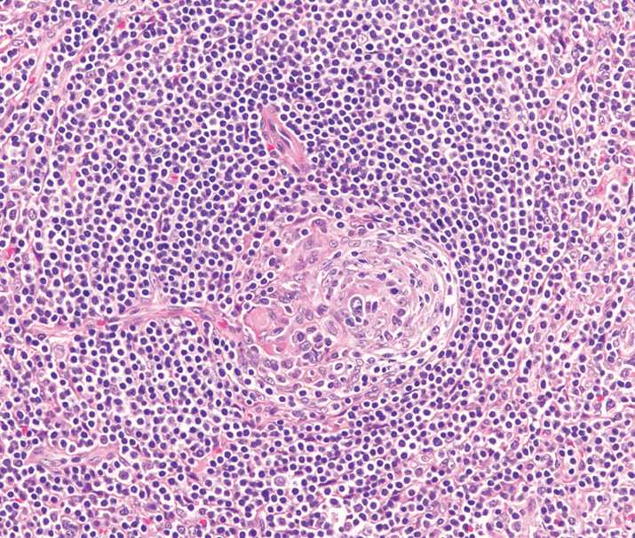
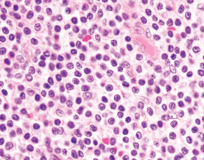
18. A young woman of Japanese descent complains of mild fever and painless lymphadenopathy. An image from her cervical lymph node excision is shown in Figure 17-3. What is the most likely diagnosis?
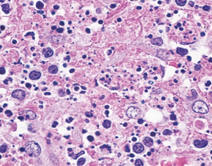
(A) Bacterial lymphadenitis
(B) Cat-scratch disease
(C) Kikuchi disease
(D) Lupus lymphadenitis
(E) Tuberculous lymphadenitis
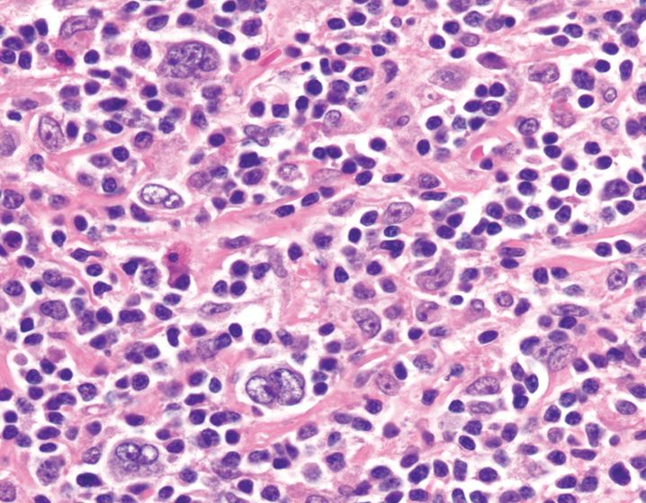
19. A 25-year-old woman with a history of fever, weight loss, and a butterfly-patterned malar rash is noted to have generalized lymphadenopathy. The clinician is concerned about an infectious process and performs a lymph node excision that is shown in Figure 17-4. Based on these clinicopathologic findings, what is the most likely cause of patient’s lymphadenopathy?
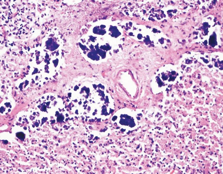
(A) Cat-scratch disease
(B) Kikuchi lymphadenitis
(C) Lupus lymphadenitis
(D) Luetic lymphadenitis
(E) Toxoplasma lymphadenitis
20. An axillary lymph node biopsy from a patient with a history of psoriasis is shown in Figure 17-5. The findings depicted in this figure are diagnostic of
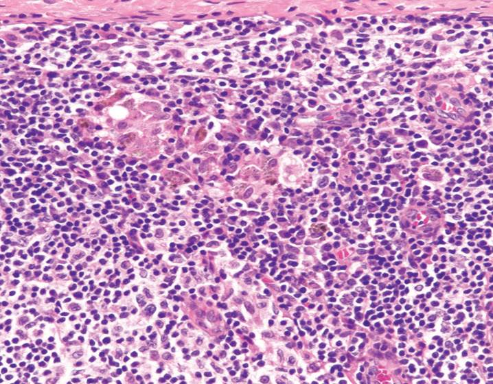
(A) Dermatopathic lymphadenitis
(B) Hodgkin lymphoma
(C) Metastatic melanoma
(D) Mycosis fungoides
(E) Toxoplasma lymphadenitis
21. All the following features are typically associated with the entity depicted in Figure 17-6, except
(A) Follicles with two or more small germinal centers
(B) Lymphocyte-depleted germinal centers
(C) PAS-positive hyaline deposits within germinal centers
(D) Sclerotic blood vessels radially penetrating the follicles
(E) Sheets of plasma cells
22. Which of the following neoplasms is not associated with Castleman’s disease?
(A) Classic Hodgkin lymphoma
(B) Diffuse large B-cell lymphoma
(C) Follicular dendritic cell sarcoma
(D) Kaposi’s sarcoma
(E) Multiple myeloma
23. Regional lymph nodes that drain tumors can be associated with all the following findings, except
(A) Desmoplastic reaction
(B) Granulomas
(C) Lymphocyte depletion
(D) Sinus histiocytosis
(E) Vascular transformation of sinuses
24. A 29-year-old patient with a history of convulsive disorder is noted to have atypical lymphocytosis in peripheral blood. A lymph node excision specimen shows relative preservation of nodal architecture and paracortical expansion by a mixed cellular infiltrate, including numerous immuno-blasts. Occasional CD30-positive large cells are also present. What is the best diagnosis?
(A) Anaplastic large cell lymphoma
(B) Angioimmunoblastic T-cell lymphoma
(C) Hodgkin lymphoma
(D) Phenytoin lymphadenitis
(E) Viral lymphadenitis
25. In which group of lymph nodes are you most likely to find lipogranulomas?
(A) Celiac
(B) Cervical
(C) Iliac
(D) Inguinal
(E) Mediastinal
26. A lymph node shows dilated sinusoids with sheets of polyethylene-containing foamy macrophages containing needle-like flakes of black material. Which of the following clinical situations can result in this finding?
(A) A patient with a history of breast implant
(B) A patient with a history of smoking
(C) A patient with a large joint prosthesis
(D) A patient with a history of intravenous drug abuse
(E) A patient with a history of rheumatoid arthritis
27. All the following features are helpful in differentiating benign glandular inclusions from metastatic adenocarcinoma, except
(A) Capsular/subcapsular location
(B) Cytokeratin CAM5.2 stain
(C) Lack of mitotic activity
(D) Lack of nuclear pleomorphism
(E) Lack of vascular/sinus invasion
28. All subtypes of Hodgkin lymphoma share the following features, except
(A) Majority of the patients are young adults
(B) Preferential involvement of cervical lymph node
(C) Presence of extensive parenchymal fibrosis
(D) Tumor cells surrounded by T cells
(E) Scattered mononuclear and multinuclear tumor cells
29. A mediastinal lymph node excision from a 40-year-old man is shown in Figure 17-7. The overall lymph node had a nodular architecture and these large cells were seen in the interfollicular region. The cells shown in this figure would express all the following immunohistochemical markers, except
(A) BCL6
(B) CD15
(C) CD20
(D) CD45
(E) CD79a
30. Based on Figure 17-8, how is this variant of Hodgkin lymphoma best classified?
(A) Lymphocyte-depleted Hodgkin lymphoma
(B) Lymphocyte-predominant Hodgkin lymphoma
(C) Mixed cellularity Hodgkin lymphoma
(D) Nodular lymphocyte-predominant Hodgkin lymphoma
(E) Nodular sclerosis Hodgkin lymphoma
31. With respect to classical Hodgkin lymphoma, what is the frequency of OCT-2 and BOB.1 expression in Hodgkin Reed-Sternberg cells?
(A) 5%
(B) 10%
(C) 50%
(D) 90%
(E) 100%
32. Within lymphoblastic lymphomas, what percentage of cases are of the B-cell phenotype?
(A) 5%
(B) 10%
(C) 50%
(D) 90%
(E) 100%
33. Which of the following molecular alterations is associated with the worst clinical prognosis in patients with B-cell acute lymphoblastic leukemia?
(A) t(4;11)
(B) t(9;22)
(C) t(12;21)
(D) Hyperdiploidy
(E) Hypodiploidy
34. What is the most common type of leukemia encountered in infants less than 1 year of age?
(A) ALL with hyperdiploidy
(B) ALL with hypodiploidy
(C) ALL with MLL gene rearrangement
(D) AML with monoblastic and monocytic features
(E) Myeloid leukemia associated with Down syndrome
35. Hyperdiploid B-cell acute lymphoblastic leukemia is most commonly associated with extra copies of the following chromosomes, except
(A) 3
(B) 4
(C) 14
(D) 21
(E) X
36. Which of the following genetic alterations in B-cell acute lymphoblastic leukemia is associated with a favorable prognosis?
(A) Hyperdiploidy
(B) Hypodiploidy
(C) t(1;19)
(D) t(4;11)
(E) t(5;14)
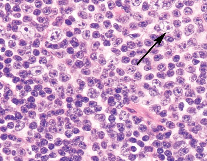
37. A lymph node excision from a 70-year-old patient with chronic lymphocytic leukemia/small lymphocytic lymphoma is shown in Figure 17-9. The larger cells present within this proliferation center are referred to as
(A) Centroblasts
(B) Centrocytes
(C) Paraimmunoblasts
(D) Plasma cells
(E) Tingible body macrophages
38. In chronic lymphocytic leukemia, all the following factors are associated with a poor prognosis, except
(A) Deletion 11q
(B) Expression of ZAP-70
(C) Expression of CD38
(D) IgHV gene mutation
(E) Rapid lymphocyte doubling time in peripheral blood
39. What percentage of peripheral blood prolymphocytes is required for a diagnosis of B-cell prolymphocytic leukemia?
(A) 5%
(B) 10%
(C) 50%
(D) 55%
(E) 90%
40. Splenic B-cell marginal zone lymphoma is associated with all of the following features, except
(A) Bone marrow may show intrasinusoidal lymphoma cells
(B) Most patients are >50 years old
(C) Peripheral blood shows presence of villous lymphocytes
(D) Tumor cells express CD5 and annexin-1
(E) Tumor involves spleen and splenic hilar lymph node
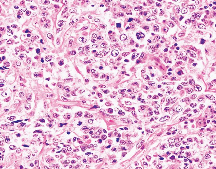
41. A splenectomy specimen from 60-year-old woman with splenomegaly, weakness, fatigue, monocytopenia, and a red pulp predominant infiltrate is shown in Figure 17-10. The cells express CD20, CD11c, CD103, CD25, and annexin-1. What is the best diagnosis?
(A) Chronic lymphocytic leukemia/small lymphocytic lymphoma
(B) Chronic myeloid leukemia
(C) Diffuse large B-cell lymphoma
(D) Hairy cell leukemia
(E) Splenic lymphoma with villous lymphocytes
42. Which hematolymphoid malignancy is most likely to be associated with IgM paraproteinemia and PAS-positive material within the lymph node sinuses?
(A) Chronic lymphocytic leukemia/small lymphocytic lymphoma
(B) Lymphoplasmacytic lymphoma
(C) Mantle cell lymphoma
(D) Marginal zone lymphoma
(E) Multiple myeloma
43. A 70-year-old woman with a history of anemia is found to have a clonal plasma cell disorder with lytic bone lesions, hypercalcemia, and renal insufficiency. The finding of elevated M-protein in this patient is due to overproduction of which of the following serum proteins?
(A) IgA
(B) IgD
(C) IgE
(D) IgG
(E) Light chain kappa
44. In addition to the lack of surface immunoglobulin expression, plasma cell myeloma cells lack expression of which of the following markers?
(A) CD19
(B) CD38
(C) CD56
(D) CD79a
(E) CD138
45. Antibiotic therapy for Helicobacter pylori (H. pylori) infection usually induces protracted remission in all cases of H. pylori-associated marginal zone lymphomas, except in those that demonstrate the following translocation
(A) t(1;14)
(B) t(3;14)
(C) t(11;18)
(D) t(14;18)
(E) +3
46. Which of the following oncogenes is most frequently rearranged in follicular lymphoma?
(A) bcl-2
(B) bcl-6
(C) myc
(D) p16
(E) p53
47. A 60-year-old man with a history of numerous gastrointestinal polyps was found to have a lymphoid neoplasm composed of CD20-positive, small-to medium-sized lymphocytes with irregular nuclear contours, and CCDN1 translocation. Which of the following choices is the most reproducible adverse prognostic parameter associated with this neoplasm?
(A) Blastoid morphology
(B) Bone marrow involvement
(C) Increased mitotic rate
(D) Increased plasma cells
(E) Peripheral blood involvement
48. What is the most common extranodal site of involvement by diffuse large B-cell lymphoma, not otherwise specified?
(A) Bone
(B) Gastrointestinal tract
(C) Skin
(D) Spleen
(E) Testis
49. A 35-year-old woman with superior vena cava syndrome is found to have an anterosuperior mediastinal mass shown in Figure 17-11. She does not have involvement of the lymph nodes or bone marrow. The tumor cells express CD20, CD30, and CD23. These findings are diagnostic of
Stay updated, free articles. Join our Telegram channel

Full access? Get Clinical Tree


