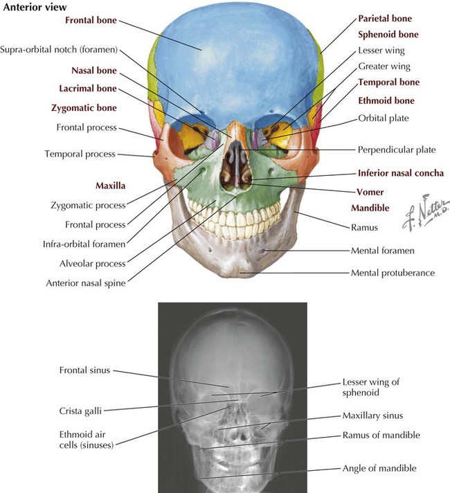
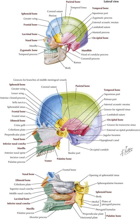
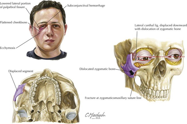
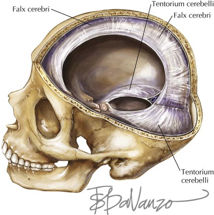
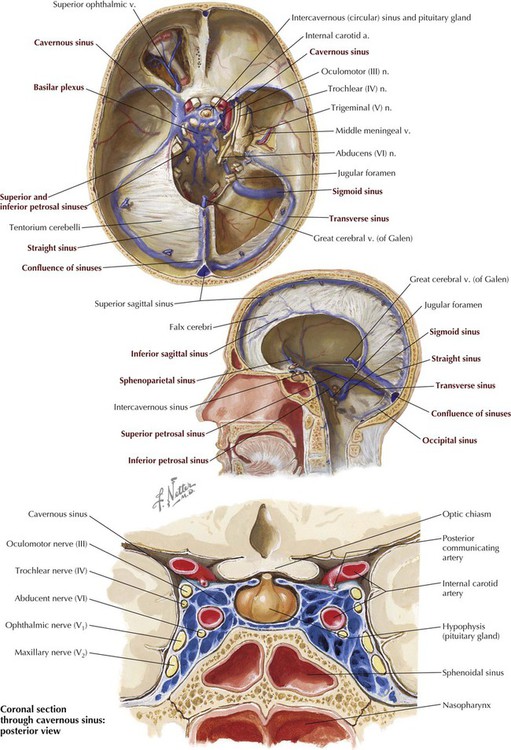
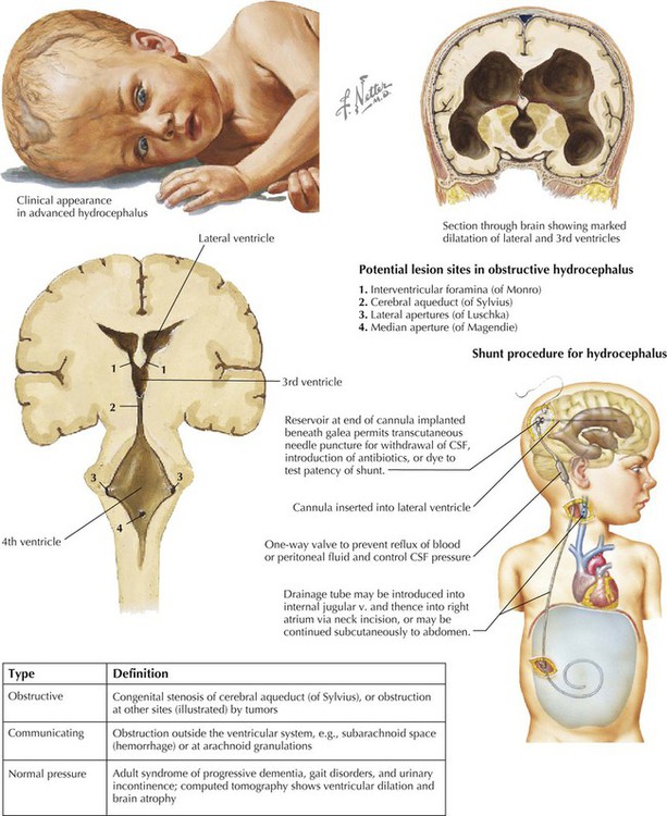
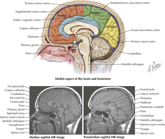
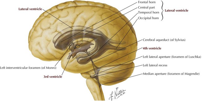
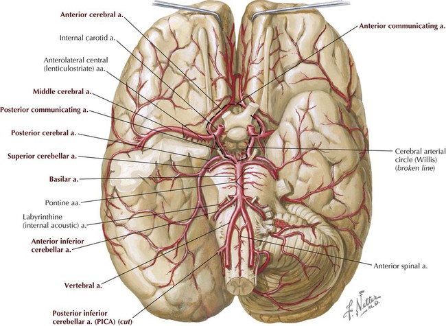
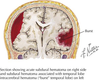
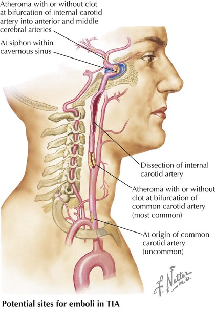
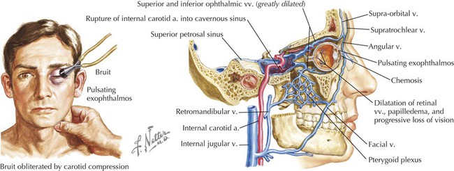
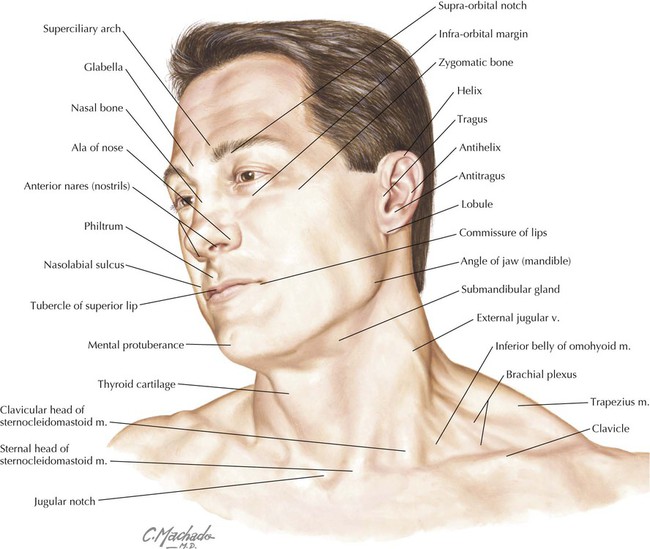
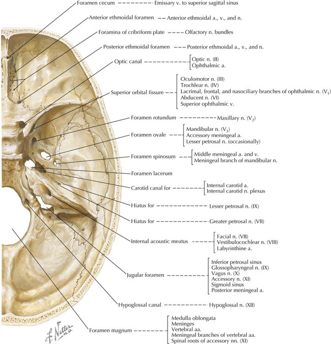
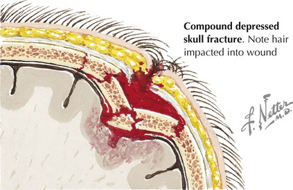
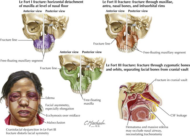
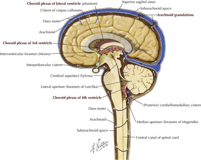
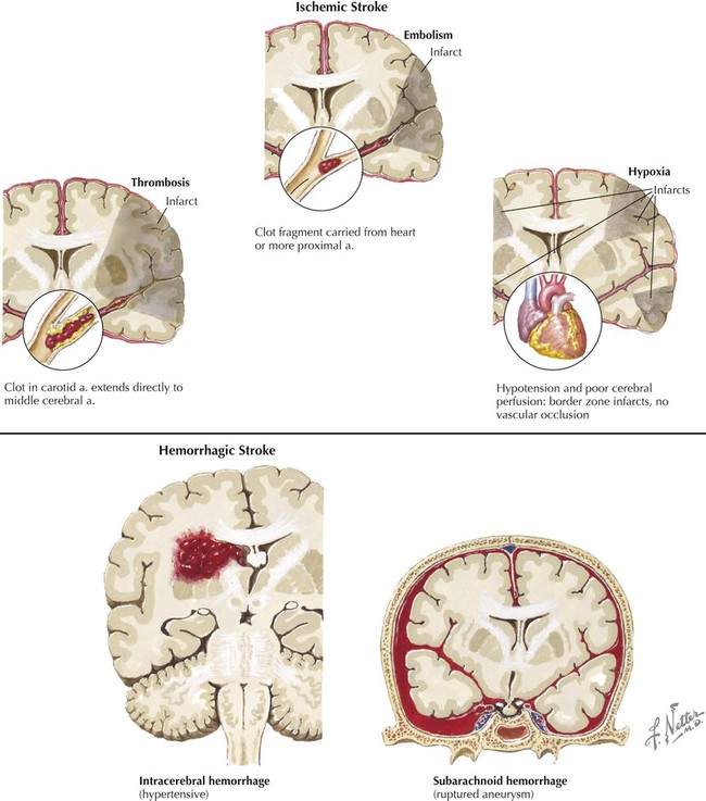
• Cranium: contains the brain and its meningeal coverings. • Orbits: contain the eye and the muscles that move the eye. • Nasal cavities and paranasal sinuses: form the uppermost part of the respiratory system. • Ears: contain the apparatus for hearing and balance. • Oral cavity: forms the proximal end of the digestive tract. • Musculofascial: superficial compartment encompassing the outer boundary of the neck. • Visceral: anterocentral compartment that contains the upper respiratory (pharynx, larynx, trachea) and gastrointestinal (GI) tract (pharynx, esophagus), and the thyroid, parathyroid, and thymus glands. • Neurovascular: two anterolateral compartments that contain the common carotid artery, internal jugular vein, and vagus nerve; called the carotid sheath. • Prevertebral: posterocentral compartment that contains the cervical vertebrae and the associated paravertebral muscles. The key surface features of the head and neck and include the following (Fig. 8-1): • Glabella: smooth prominence on the frontal bone above the root of the nose. • Zygomatic bone: the cheekbone, which protrudes below the orbit and is vulnerable to fractures from facial trauma. • Ear (auricle or pinna): skin-covered elastic cartilage with several consistent ridges, including the helix, antihelix, tragus, antitragus, and lobule. • Philtrum: midline infranasal depression of the upper lip. • Nasolabial sulcus: line between the nose and the corner of the lip. • Thyroid cartilage: the laryngeal prominence (“Adam’s apple”). • Jugular (suprasternal) notch: midline depression between the two sternal heads of the sternocleidomastoid muscle. The skull is composed of 22 bones (see Chapter 1). Eight of these bones form the cranium (neurocranium, which contains the brain and meninges), and 14 of these form the face (viscerocranium). There are seven associated bones: the auditory ossicles (three in each middle ear) and the unpaired hyoid bone (Fig. 8-2 and Table 8-1). Using your atlas and dry bone specimens, note the complexity of the maxillary, temporal, and sphenoid bones. These bones are in close association with many of the cranial nerves and encase portions of many of our special senses—balance, hearing, smell, sight, and even taste—as the maxillae form a portion of the oral cavity. TABLE 8-1 Other features of the skull are noted as we review each region of the head. However, general external features include the following (Figs. 8-2 and 8-3): • Coronal suture: region between the frontal and two parietal bones. • Sagittal suture: region between the two parietal bones. • Lambdoid suture: region between the occipital bone and the two parietal bones. • Nasion: point at which the frontal and nasal bones meet. • Bregma: point at which coronal and sagittal sutures meet. • Lambda: point at which sagittal and lambdoid sutures meet. • Pterion: point at which frontal, sphenoid, temporal, and parietal bones meet; the middle meningeal artery lies beneath this region. • Asterion: point at which temporal, parietal, and occipital bones meet. The cranial base is the floor of the neurocranium, which supports the brain, and is divided into the following three cranial fossae: (Fig. 8-4): • Anterior: the roof of the orbits; accommodates the frontal lobes of the brain. • Middle: accommodates the temporal lobes of the brain. • Posterior: accommodates the cerebellum, pons, and medulla oblongata of the brain. Each fossa has numerous foramina (openings) for structures to pass in or out of the neurocranium. Clinical Focus 8-1 Skull Fractures Skull fractures may be classified as follows: • Linear: presents with a distinct fracture line. • Comminuted: presents with multiple fragments (depressed if driven inward; can compress or tear the underlying dura mater). • Diastasis: fracture along a suture line. Clinical Focus 8-3 Midface Fractures • Le Fort I: horizontal detachment of the maxilla at the level of the nasal floor (ant.-post. views). • Le Fort II: pyramidal fracture that includes both maxillae and nasal bones, medial portions of both maxillary antra, infra-orbital rims, orbits, and orbital floors (ant.-post. views). • Le Fort III: includes Le Fort II and a fracture of both zygomatic bones; may cause airway problems, nasolacrimal apparatus obstruction, and cerebrospinal fluid leakage (ant.-post. views). The brain and spinal cord are surrounded by three membranous connective tissue layers called the meninges, which include the following (Fig. 8-5): • Dura mater: thick outermost meningeal layer that is richly innervated by sensory nerve fibers. • Arachnoid mater: fine, weblike avascular membrane directly beneath the dural surface; the space between the arachnoid and the underlying pia is called the subarachnoid space and contains cerebrospinal fluid, which bathes and protects the central nervous system (CNS). • Pia mater: delicate membrane of connective tissue that intimately envelops the brain and spinal cord. The dura mater is richly innervated by meningeal sensory branches of the trigeminal nerve (fifth cranial nerve, CN V); the vagus nerve (CN X), specifically to the posterior cranial fossa; and the upper cervical nerves. A portion of the dura in the posterior cranial fossa also may receive some innervation from the glossopharyngeal nerve (CN IX) and hypoglossal nerve (CN XII). The arachnoid and pia mater lack sensory innervation. The periosteal dura and meningeal dura separate to form thick connective tissue folds or layers that separate various brain regions and lobes (Figs. 8-5, 8-6, and 8-7): • Falx cerebri: double layer of meningeal dura between the cerebral hemispheres. • Falx cerebelli: sickle-shaped layer of meningeal dura that projects between the two cerebellar hemispheres. • Tentorium cerebelli: fold of meningeal dura that covers the cerebellum and supports the occipital lobes of the cerebral hemispheres. • Diaphragma sellae: horizontal shelf of meningeal dura that forms the roof of the sella turcica covering the pituitary gland; the infundibulum passes through this dural shelf to connect the hypothalamus with the pituitary gland. The dura also separates to form several large endothelial-lined venous channels between its periosteal and meningeal layers—superior and inferior sagittal, straight, confluence of sinuses, transverse, sigmoid, and cavernous sinuses—and several smaller dural sinuses (Table 8-2 and Fig. 8-7). These dural venous sinuses drain blood from the brain, largely posteriorly, then into the internal jugular veins. These sinuses lack valves, however, so the direction of blood flow through the sinuses is pressure dependent. Of particular importance is the cavernous venous sinus, which lies on either side of the sella turcica and has an anatomical relationship with the internal carotid artery and several cranial nerves, including III, IV, V1, V2, and VI. Injury or inflammation in this region can affect all these important structures. Also, the optic chiasm lies just above this area, so CN II may be involved in any superior expansion of the cavernous sinus (e.g., pituitary tumor). TABLE 8-2 The subarachnoid space (between the arachnoid and pia mater) contains cerebrospinal fluid (CSF), which performs the following functions (Figs. 8-5 and 8-8): • Supports and cushions the spinal cord and brain. • Fulfills some functions normally provided by the lymphatic system. • Occupies a volume of about 150 mL in the subarachnoid space. • Is produced by choroid plexuses in the brain’s ventricles. • Is produced at a rate of about 500 to 700 mL/day. • Is reabsorbed largely by arachnoid granulations and by small venules along the length of the spinal cord. The arachnoid granulations absorb most of the CSF and deliver it to the dural venous sinuses (see Figs. 8-5 and 8-8). These granulations are composed of convoluted aggregations of arachnoid that extend as “tufts” into the superior sagittal sinus and function as one-way valves for the clearance of CSF; the CSF crosses into the venous sinus, but venous blood cannot enter the subarachnoid space. Small, microscopic arachnoid cell herniations also occur along the spinal cord, where CSF (which circulates at a higher pressure than venous blood) is delivered directly into small spinal cord veins. The most notable feature of the human brain is its large cerebral hemispheres (Fig. 8-9). Several circumscribed regions of the cerebral cortex are associated with specific functions, and key surface landmarks of the typical human cerebrum are used to divide the brain into lobes: four or five, depending on classification, with the fifth either the insula or the limbic lobe. The lobes and their functions are as follows: • Frontal: mediates precise voluntary motor control, learned motor skills, planned movement, eye movement, expressive speech, personality, working memory, complex problem solving, emotions, judgment, socialization, olfaction, and drive. • Parietal: affects sensory input, spatial discrimination, sensory representation and integration, taste, and receptive speech. • Occipital: affects visual input and processing. • Temporal: mediates auditory input and auditory memory integration, spoken language (dominant side), and body language (nondominant side). • Insula: a fifth deep lobe that lies medial to the temporal lobe (sometimes included as part of temporal lobe); influences vestibular function, some language, perception of visceral sensations (e.g., upset stomach), emotions, and limbic functions. • Limbic: also sometimes considered a fifth medial lobe (cingulate cortex); influences emotions and some autonomic functions. Other key areas of the brain include the following components (Fig. 8-9): • Thalamus: gateway to the cortex; simplistically functions as an “executive secretary” to the cortex (relay center between cortical and subcortical areas). • Cerebellum: coordinates smooth motor activities, and processes muscle position; possible role in behavior and cognition. • Brainstem: includes the midbrain, pons, and medulla oblongata; conveys motor and sensory information from the body and autonomic and motor information from higher centers to peripheral targets. Internally, the brain contains four ventricles, two lateral ventricles, and a central third and fourth ventricle (Fig. 8-10). Cerebrospinal fluid, produced by the choroid plexus (see Fig. 8-5), circulates through these ventricles and then enters the subarachnoid space through two lateral apertures (foramina of Luschka) or a median aperture (foramen of Magendie) in the fourth ventricle. Arteries supplying the brain arise largely from the following two pairs of arteries (Fig. 8-11 and Table 8-3): TABLE 8-3 • Vertebrals: arise from the subclavian artery, ascend through the transverse foramina of the C1-C6 vertebrae, and enter the foramen magnum of the skull. • Internal carotids: arise from the common carotid in the neck, ascend in the neck, enter the carotid canal, and traverse the foramen lacerum to terminate as the middle and anterior cerebral arteries, which anastomose with the arterial circle of Willis. The vertebral arteries give rise to the anterior and posterior spinal arteries (a portion of the supply to the spinal cord) and the posterior inferior cerebellar arteries, and then join at about the level of the junction between the medulla and pons to form the basilar artery (Fig. 8-11). The internal carotid arteries each give rise to an ophthalmic artery, a posterior communicating artery, a middle cerebral artery, and an anterior cerebral artery. Table 8-3 summarizes the brain regions supplied by these vessels and their major branches. Clinical Focus 8-10 Stroke • Ischemic (80%): infarction; thrombotic or embolic, resulting from atherosclerosis of the extracranial (usually carotid) and intracranial arteries or from underlying heart disease. • Hemorrhagic: occurs when a cerebral vessel weakens and ruptures (subarachnoid or intracerebral hemorrhage), which causes intracranial bleeding, usually affecting a larger brain area.
Head and Neck
1 Introduction
2 Surface Anatomy
3 Skull
BONE
DESCRIPTION
Frontal
Forms forehead, is thicker anteriorly, contains frontal sinuses
Nasal
Paired bones that form the root of the nose
Lacrimal
Small, paired bones that form part of the anteromedial wall of the orbit and contain the lacrimal sac
Zygomatic
Paired cheekbones that form the inferolateral rim of the orbit and are frequently fractured by blunt trauma
Maxilla
Paired bones that form part of the cheek and contain 16 maxillary teeth
Mandible
Lower jaw bone that contains 16 mandibular teeth
Parietal
Forms the superolateral portion of the neurocranium
Temporal
Paired bones that form the lower portion of the lateral neurocranium and contain the middle and inner ear cavities, and the vestibular system for balance
Sphenoid
Complex bone composed of a central body, and greater and lesser wings
Occipital
Forms the inferoposterior portion of the neurocranium
Ethmoid
Forms the ethmoid sinuses, and contributes to the medial, lateral, and superior walls of the nasal cavity
Inferior concha
Paired bones of the lateral nasal wall that form the inferior nasal concha
Vomer
Forms the lower part of the nasal septum
Palatine
Contributes to the lateral nasal wall, a small part of the nasal septum, and the hard palate
Cranial Fossae
4 Brain
Meninges
Dural Venous Sinuses
SINUS
CHARACTERISTICS
Superior sagittal
Midline sinus along the convex superior border of the falx cerebri
Inferior sagittal
Midline sinus along the inferior free edge of the falx cerebri and joined by the great cerebral vein (of Galen)
Straight
Runs in the attachment of the falx cerebri and the tentorium cerebelli, and is formed by the inferior sagittal sinus and great cerebral vein
Confluence of sinuses
Meeting of superior and inferior sagittal sinuses, the straight sinus, and the occipital sinus
Transverse
Extends from the confluence of sinuses along the lateral edge of the tentorium cerebelli
Sigmoid
Continuation of the transverse sinus that passes inferomedially in an S-shaped pathway to the jugular foramen (becomes internal jugular vein)
Occipital
Runs in the falx cerebelli to the confluence of sinuses
Basilar
Network of venous channels on basilar part of the occipital bone, with connections to the petrosal sinuses; drains into vertebral venous plexus
Cavernous
Lies between dural layers on each side of the sella turcica; connects to the superior ophthalmic veins, pterygoid plexus of veins, sphenoparietal sinuses, petrosal sinuses, and basilar sinus
Sphenoparietal
Runs along the posterior edge of the lesser wing of the sphenoid bone and drains into the cavernous sinus
Emissary veins
Small veins connect the dural sinuses with the diploic veins in the bony skull, which are connected to scalp veins
Subarachnoid Space

Gross Anatomy of the Brain
Blood Supply to the Brain
ARTERY
COURSE AND STRUCTURES SUPPLIED
Vertebral
From subclavian artery; supplies cerebellum
Posterior inferior cerebellar
From vertebral artery; supplies the posteroinferior cerebellum
Basilar
From both vertebrals; supplies brainstem, cerebellum, and cerebrum
Anterior inferior cerebellar
From basilar; supplies inferior cerebellum
Superior cerebellar
From basilar; supplies superior cerebellum
Posterior cerebral
From basilar; supplies inferior cerebrum and occipital lobe
Posterior communicating
Cerebral arterial circle (of Willis)
Internal carotid (IC)
From common carotid; supplies cerebral lobes and eye
Middle cerebral
From IC; supplies lateral aspect of cerebral hemispheres
Anterior communicating
Cerebral arterial circle (of Willis)
Anterior cerebral
From IC; supplies cerebral hemispheres (except occipital lobe)
Head and Neck