Halo Nevi
David Cassarino, MD, PhD
Key Facts
Terminology
Nevus with clinically depigmented halo surrounding pigmented area
Clinical Issues
Usually young patients (children and young adults)
In older patients, should raise concern for melanoma
Microscopic Pathology
Nevus associated with dense inflammatory infiltrate
Infiltrate typically shows a lichenoid pattern in dermis
Nests predominate in early lesions, single cells later
Melanocytic markers may be useful to confirm presence of melanocytes
Reactive atypia may be present
Top Differential Diagnoses
Melanoma
Myerson nevus (eczematous nevus)
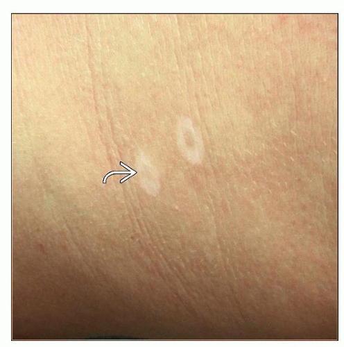 Two halo nevi seen on the back of a young adult are oval, well demarcated, and depigmented (skin colored or paler). With time, the white area may replace the nevus entirely  . . |
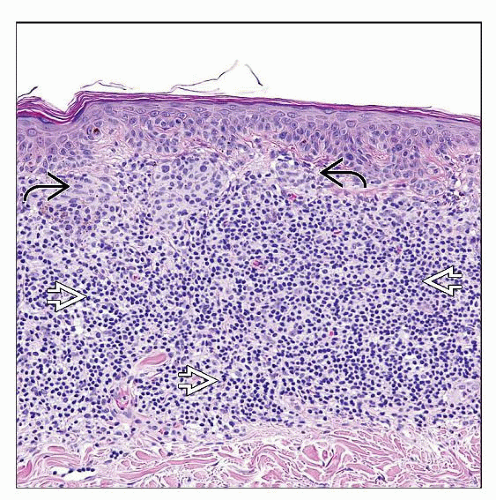 Halo nevus is characterized by a dense, band-like lymphohistiocytic infiltrate in the dermis  . Junctional and superficial nests of melanocytes can be appreciated upon close inspection . Junctional and superficial nests of melanocytes can be appreciated upon close inspection  . . |
TERMINOLOGY
Synonyms
Sutton nevus
Nevus depigmentosa centrifugum
Definitions
Nevus with clinically depigmented halo surrounding pigmented area
Dense inflammatory infiltrate typically present
Histologically heavily inflamed nevi that lack a clinical halo may be said to show “halo reaction/phenomenon,” but they are not true halo nevi
ETIOLOGY/PATHOGENESIS
Inflammatory Process
Thought to be a reaction to melanocytic antigens
Stay updated, free articles. Join our Telegram channel

Full access? Get Clinical Tree


