H


H


Halitosis

Halitosis describes any breath odor that’s unpleasant, disagreeable, or offensive. This common sign is usually easy to detect, but an embarrassed patient may take measures to hide it. The patient may be unaware that he has halitosis, even though he may complain of a bad taste in his mouth, or he may believe that he has halitosis but that no one else can detect it (psychogenic halitosis).

Certain types of halitosis characterize specific disorders—for example, a fruity breath odor typifies ketoacidosis. (See “Breath with ammonia odor,” page 120; “Breath with fecal odor,” page 121; “Breath with fruity odor,” page 123; and “Fetor hepaticus,” page 297.) Other types of halitosis include putrid, foul, fetid, and musty breath odors.

Halitosis may result from a disorder of the oral cavity, nasal passages, sinuses, respiratory tract, or esophageal diverticula. It may also stem from a GI disorder associated with belching, regurgitation, or vomiting, or it may be an adverse effect of an oral or inhaled drug.

Other causes of halitosis include cigarette smoking, ingestion of alcohol and certain foods (such as garlic and onions), and poor oral hygiene—especially in patients with an orthodontic device, dentures, or dental caries. In addition, offensive skin odors—for example, from foot perspiration—may be absorbed locally and later expelled by the lungs, resulting in halitosis.


HISTORY AND PHYSICAL EXAMINATION

If you detect halitosis, try to characterize the odor. Does it smell fruity, fecal, or musty? If the patient is aware of it, find out how long he has had it. Does he also have a bad taste in his mouth? Does he have difficulty swallowing or chewing? Does he have reflux or regurgitation? Does he have pain or tenderness? Ask the patient if he has a problem with flatus. Also ask him to describe the frequency of his bowel movements and the size and consistency of his stools.

Find out if the patient smokes or chews tobacco. Have him describe his diet and daily oral hygiene. Does he wear dentures? Complete the history by asking about chronic disorders and recent respiratory tract infection. If the patient reports a cough, find out if it’s productive.

Begin the physical examination by examining the patient’s mouth, throat, and nose. Look for lesions, bleeding, drainage, obstruction, and signs of infection, such as redness and swelling. Check for tenderness by percussing and palpating over the sinuses. Then auscultate the lungs for abnormal breath sounds. Auscultate the abdomen for bowel sounds, and percuss it, noting any tympany. Finally, take vital signs.


MEDICAL CAUSES

Bowel obstruction. Halitosis is a late sign in both small- and large-bowel obstructions, resulting from vomiting of bilious and later fecal material. Other findings in a small-bowel obstruction include constipation, abdominal
distention, and intermittent periumbilical cramping pain. In a large-bowel obstruction, abdominal pain is milder and more constant than that associated with a small-bowel obstruction and is usually located lower in the abdomen.

Bronchiectasis. Bronchiectasis usually produces foul or putrid halitosis, but some patients may have a sickeningly sweet breath odor. The patient typically also has a chronic productive cough with copious, foul-smelling, mucopurulent sputum. The cough is aggravated by lying down and is most productive in the morning. Associated findings commonly include exertional dyspnea, fatigue, malaise, weakness, and weight loss. Auscultation reveals coarse or moist crackles over the affected lung areas during inspiration. Digital clubbing is a late sign.

Common cold. A musty breath odor may accompany a common cold, which usually also causes a dry, hacking cough with sore throat, sneezing, nasal congestion, rhinorrhea, headache, malaise, fatigue, arthralgia, and myalgia.

Esophageal cancer. In esophageal cancer, halitosis may accompany classic findings of dysphagia, hoarseness, chest pain, and weight loss. Nocturnal regurgitation and cachexia are late signs.

Gastric cancer. Halitosis is a late sign in gastric cancer. Accompanying findings include chronic dyspepsia unrelieved by antacids, a vague feeling of fullness, nausea, anorexia, fatigue, pallor, weakness, altered bowel habits, weight loss, and muscle wasting. Hematemesis and melena are signs of associated gastric bleeding.

Gastrocolic fistula. In this disorder, fecal vomiting is responsible for fecal breath odor, which is typically preceded by intermittent diarrhea.

Gingivitis. Characterized by red, edematous gums, gingivitis may also cause halitosis. The gingivae between the teeth become bulbous and bleed easily with slight trauma.

Acute necrotizing ulcerative gingivitis also causes fetid breath, a bad taste in the mouth, and ulcers—especially between the teeth—that may become covered with a gray exudate. Severe ulceration may occur with fever, cervical adenopathy, headache, and malaise.

Hepatic encephalopathy. A characteristic late sign of hepatic encephalopathy is fetor hepaticus, a musty, sweet, or mousy (new-mown hay) breath odor. Other late effects include coma, asterixis (flapping tremor), and hyperactive deep tendon reflexes.

Ketoacidosis. Alcohol-induced, diabetic, and starvation forms of ketoacidosis produce a fruity breath odor. Alcohol-induced ketoacidosis is usually seen in poorly nourished alcoholics who have eaten very little over several days. Symptoms include sudden Kussmaul’s respirations with vomiting for several days, light dehydration, abdominal pain and distention, and absent bowel sounds. The patient is alert and has a normal or slightly decreased blood glucose level.

Life-threatening diabetic ketoacidosis produces a rapid, thready pulse; marked hypovolemia; nausea and vomiting; and, in its early stages, the triad of polydipsia, polyphagia, and polyuria.

Also life-threatening, starvation ketoacidosis produces Kussmaul’s respirations; weight loss; bradycardia; dry, scaly skin; sore tongue; muscle and tissue wasting; abdominal distention; and signs of dehydration, such as oliguria and poor skin turgor.

Other common effects of diabetic and starvation ketoacidosis include orthostatic hypotension, generalized weakness, anorexia, abdominal pain, and altered level of consciousness.

Lung abscess. Lung abscess typically causes putrid halitosis, but its cardinal sign is a productive cough with copious, purulent, often bloody sputum. Other findings include fever with chills, dyspnea, headache, anorexia, weight loss, malaise, pleuritic chest pain, asymmetrical chest movement, and temporary clubbing.

Necrotizing ulcerative mucositis (acute). A strong, putrid breath odor is characteristic of this uncommon disorder, which initially causes slight cheek inflammation that’s rapidly followed by tooth loss and extensive bone sloughing in the mandible or maxilla.

Ozena. This severe, chronic form of rhinitis causes a musty or fetid breath odor as well as thick green mucus and progressive anosmia.

Periodontal disease. Periodontal disease causes halitosis and an unpleasant taste. Typically, the patient’s gums bleed spontaneously or with slight trauma and are marked by pus-filled pockets around the teeth. Related findings include facial pain, headache, and loose teeth covered by calculi and plaque.

Pharyngitis (gangrenous). Halitosis is a chief sign of gangrenous pharyngitis. The patient also complains of a foul taste in the mouth, an extremely sore throat, and a choking sensation. Examination reveals a swollen, red, ulcerated pharynx, possibly with a grayish
membrane. Fever and cervical lymphadenopathy are also common.

Renal failure (chronic). Renal failure produces a urinous or ammonia breath odor. Among its widespread effects are anemia, emotional lability, lethargy, irritability, decreased mental acuity, coarse muscular twitching, peripheral neuropathies, muscle wasting, anorexia, signs of GI bleeding, ecchymosis, yellowbrown or bronze skin, pruritus, anuria, and increased blood pressure.

Sinusitis. Acute sinusitis causes a purulent nasal discharge that leads to halitosis. Besides a characteristic postnasal drip, the patient may exhibit nasal congestion, sore throat, cough, malaise, headache, facial pain and tenderness, and fever. Chronic sinusitis causes a continuous mucopurulent discharge that leads to a musty breath odor, postnasal drip, nasal congestion, and a chronic nonproductive cough.

Zenker’s diverticulum. This esophageal disorder causes halitosis and a bad taste in the mouth associated with regurgitation. The patient may also report a chronic cough that’s most pronounced at night, hoarseness, odynophagia, neck pain, and “gurgling” sounds in the throat when he swallows liquids.


OTHER CAUSES

Drugs. Drugs that can cause halitosis include triamterene, inhaled anesthetics, and any drugs known to cause metabolic acidosis such as nitroprusside.

image Some herbal medicines, such as garlic, may cause halitosis.


SPECIAL CONSIDERATIONS

If examination of the mouth and sinuses doesn’t reveal the cause of halitosis, prepare the patient for upper GI and chest X-rays or endoscopy.


PEDIATRIC POINTERS

In children, halitosis commonly results from physiologic causes, such as continual mouth breathing and thumb or blanket sucking. Phenylketonuria—a metabolic disorder that affects infants—may produce a musty or mousy breath odor.


GERIATRIC POINTERS

Extensive dental caries, mouth dryness, and poor oral hygiene can cause halitosis in elderly patients.


PATIENT COUNSELING

To help control halitosis, encourage good oral hygiene. If halitosis is drug induced, reassure the patient that it will disappear as soon as his body completely eliminates the drug.


Halo vision

Halo vision refers to seeing rainbowlike colored rings around lights or bright objects. The rainbowlike effect can be explained by this physical principle: As light passes through water (in the eye, through tears or the cells of various anteretinal media), it breaks up into spectral colors.

Halo vision usually develops suddenly; its duration depends on the causative disorder. This symptom may occur in disorders associated with excessive tearing and corneal epithelial edema. Among these causes, the most common and significant is acute angle-closure glaucoma, which can lead to blindness. In this ophthalmic emergency, increased intraocular pressure (IOP) forces fluid into corneal tissues anterior to Bowman’s membrane, causing edema. Halo vision is also an early symptom of cataracts, resulting from dispersion of light by abnormal lens opacity.

Nonpathologic causes of excessive tearing associated with halo vision include poorly fitted or overworn contact lenses, emotional extremes, and exposure to intense light, as in snow blindness.


HISTORY AND PHYSICAL EXAMINATION

First, ask the patient how long he has been seeing halos around lights and when he usually sees them. Patients with glaucoma usually see halos in the morning, when IOP is most elevated. Ask the patient if light bothers his eyes. Does he have eye pain? If so, have him describe it. Remember that halos associated with excruciating eye pain or a severe headache may point to acute angle-closure glaucoma, an ocular emergency. Note a history of glaucoma or cataracts.

Next, examine the patient’s eyes, noting conjunctival injection, excessive tearing, and lens changes. Examine pupil size, shape, and response to light. Then test visual acuity by performing an ophthalmoscopic examination.


MEDICAL CAUSES

Cataract. Halo vision may be an early symptom of painless, progressive cataract formation.
The glare of headlights may blind the patient, making nighttime driving impossible. Other features include blurred vision, impaired visual acuity, and lens opacity, all of which develop gradually.

Corneal endothelial dystrophy. Typically, halo vision is a late symptom of this disorder, which may also cause impaired visual acuity.

Glaucoma. Halo vision characterizes all types of glaucoma. Acute angle-closure glaucoma —an ophthalmic emergency—also causes blurred vision, followed by a severe headache or excruciating pain in and around the affected eye. Examination reveals a moderately dilated fixed pupil that doesn’t respond to light, conjunctival injection, a cloudy cornea, impaired visual acuity and, possibly, nausea and vomiting.

Chronic angle-closure glaucoma usually produces no symptoms until pain and blindness occur in advanced disease. Sometimes, halos and blurred vision develop slowly.

In chronic open-angle glaucoma, halo vision is a late symptom that’s accompanied by mild eye ache, peripheral vision loss, and impaired visual acuity.


SPECIAL CONSIDERATIONS

To help minimize halo vision, remind the patient not to look directly at bright lights.


PEDIATRIC POINTERS

Halo vision in a child usually results from congenital cataracts or glaucoma. In a young child, limited verbal ability may make halo vision difficult to assess.


GERIATRIC POINTERS

Primary glaucoma, the most common cause of halo vision, is more common in older patients.


Headache

The most common neurologic symptom, headaches may be localized or generalized, producing mild to severe pain. About 90% of all headaches are benign and can be described as vascular, muscle-contraction, or a combination of both. (See Comparing benign headaches.) Occasionally, though, headaches indicate a severe neurologic disorder associated with intracranial inflammation, increased intracranial pressure (ICP), or meningeal irritation. They may also result from an ocular or sinus disorder, tests, drugs, or other treatments.

Other causes of headache include fever, eyestrain, dehydration, and systemic febrile illnesses. Headaches may occur in certain metabolic disturbances—such as hypoxemia, hypercapnia, hyperglycemia, and hypoglycemia—but they aren’t a diagnostic or prominent symptom in these disorders. Some individuals get headaches after seizures or from coughing, sneezing, heavy lifting, or stooping.


HISTORY AND PHYSICAL EXAMINATION

If the patient reports a headache, ask him to describe its characteristics and location. How often does he get a headache? How long does a typical headache last? Try to identify precipitating factors, such as eating certain foods or exposure to bright lights. Ask what helps to relieve the headache. Is the patient under stress? Has he had trouble sleeping?

Take a drug and alcohol history, and ask about head trauma within the last 4 weeks. Has the patient recently experienced nausea, vomiting, photophobia, or visual changes? Does he feel drowsy, confused, or dizzy? Has he recently developed seizures, or does he have a history of seizures?

Begin the physical examination by evaluating the patient’s level of consciousness (LOC). Then check his vital signs. Be alert for signs of increased ICP—widened pulse pressure, bradycardia, altered respiratory pattern, and increased blood pressure. Check pupil size and response to light, and note any neck stiffness. (See Differential diagnosis: Headache, pages 344 and 345.)


MEDICAL CAUSES

Anthrax, cutaneous. Along with a macular or papular lesion that develops into a vesicle and finally a painless ulcer, this disorder may produce a headache, lymphadenopathy, fever, and malaise.

Brain abscess. In this disorder, the headache is localized to the abscess site; it usually intensifies over a few days and is aggravated by straining. Accompanying the headache may be nausea, vomiting, and focal or generalized seizures. The patient’s LOC varies from drowsiness to deep stupor. Depending on the abscess site, associated signs and symptoms may include aphasia, impaired visual acuity, hemiparesis, ataxia, tremors, and personality changes. Signs of infection, such as fever and pallor, usually develop late; however, if the

abscess remains encapsulated, these signs may not appear.


Brain tumor. Initially, a tumor causes a localized headache near the tumor site; as the tumor grows, the headache eventually becomes generalized. The pain is usually intermittent, deep seated, and dull and is most intense in the morning. It’s aggravated by coughing, stooping, Valsalva’s maneuver, and changes in head position, and it’s relieved by sitting and rest.
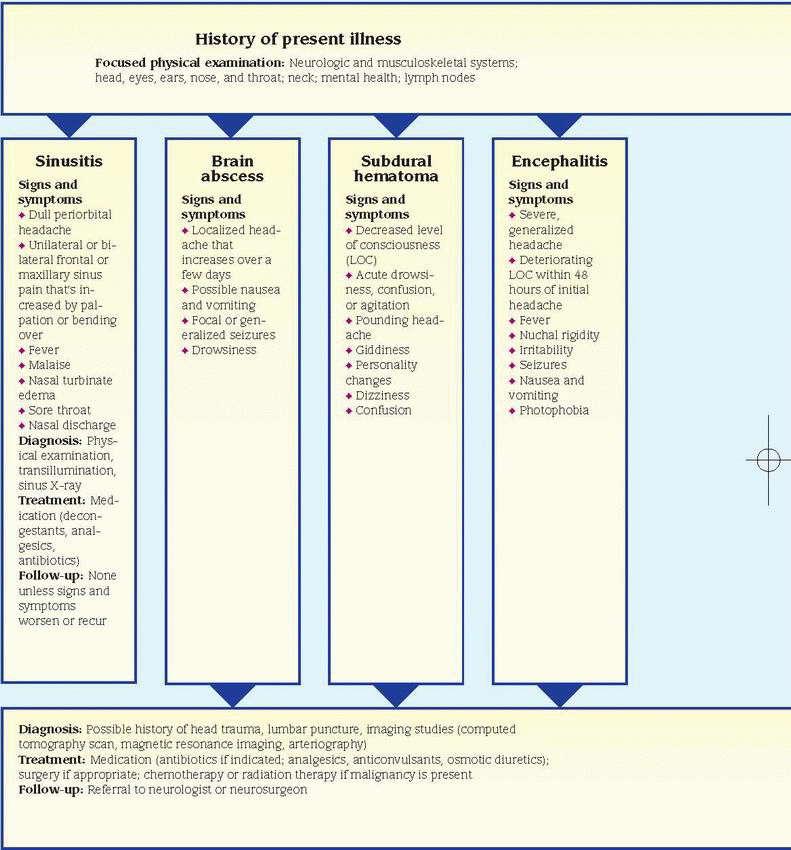
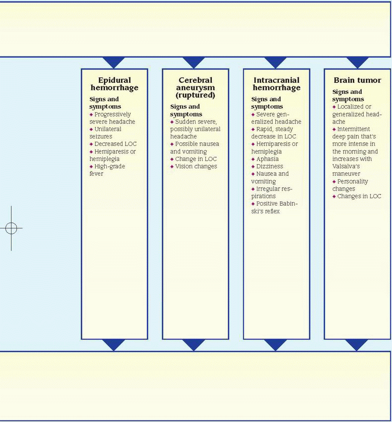
Associated signs and symptoms include personality changes, altered LOC, motor and sensory dysfunction, and eventually signs of increased ICP, such as vomiting, increased systolic blood pressure, and widened pulse pressure.

Cerebral aneurysm (ruptured). Cerebral aneurysm is a life-threatening disorder that’s characterized by a sudden excruciating headache, which may be unilateral and usually peaks within minutes of the rupture. The patient
may lose consciousness immediately or display a variably altered LOC. Depending on the severity and location of the bleeding, he may also exhibit nausea and vomiting; signs and symptoms of meningeal irritation, such as nuchal rigidity and blurred vision; hemiparesis; and other features.

Ebola virus. A sudden headache commonly occurs on the 5th day of this deadly illness. Additionally, the patient has a history of malaise, myalgia, high fever, diarrhea, abdominal pain, dehydration, and lethargy. A maculopapular rash develops between the 5th and 7th days of the illness. Other possible findings include pleuritic chest pain; a dry, hacking cough; pronounced pharyngitis; hematemesis; melena; and bleeding from the nose, gums, and vagina. Death usually occurs in the 2nd week of the illness, preceded by massive blood loss and shock.

Encephalitis. A severe, generalized headache is characteristic with this disorder. Within 48 hours, the patient’s LOC typically deteriorates —perhaps from lethargy to coma. Associated signs and symptoms include fever, nuchal rigidity, irritability, seizures, nausea and vomiting, photophobia, cranial nerve palsies such as ptosis, and focal neurologic deficits, such as hemiparesis and hemiplegia.

Epidural hemorrhage (acute). Head trauma and a sudden, brief loss of consciousness usually precede this hemorrhage, which causes a progressively severe headache that’s accompanied by nausea and vomiting, bladder distention, confusion, and then a rapid decrease in LOC. Other signs and symptoms include unilateral seizures, hemiparesis, hemiplegia, high fever, decreased pulse rate and bounding pulse, widened pulse pressure, increased blood pressure, a positive Babinski’s reflex, and decerebrate posture.

If the patient slips into a coma, his respirations deepen and become stertorous, then shallow and irregular, and eventually cease. Pupil dilation may occur on the same side as the hemorrhage.

Glaucoma, acute angle-closure. This type of glaucoma is an ophthalmic emergency that may cause an excruciating headache as well as acute eye pain, blurred vision, halo vision, nausea, and vomiting. Assessment reveals conjunctival injection, a cloudy cornea, and a moderately dilated, fixed pupil.

Hantavirus pulmonary syndrome. Noncardiogenic pulmonary edema distinguishes this viral disease, which was first reported in the United States in 1993. Common reasons for seeking treatment include flulike signs and symptoms—headache, myalgia, fever, nausea, vomiting, and a cough—followed by respiratory distress. Fever, hypoxia, and (in some patients) serious hypotension typify the hospital course. Other signs and symptoms include a rising respiratory rate (28 breaths/minute or more) and an increased heart rate (120 beats/minute or more).

Hypertension. This disorder may cause a slightly throbbing occipital headache on awakening that decreases in severity during the day. However, if the patient’s diastolic blood pressure exceeds 120 mm Hg, the headache remains constant. Associated signs and symptoms include an atrial gallop, restlessness, confusion, nausea and vomiting, blurred vision, seizures, and altered LOC.

Influenza. A severe generalized or frontal headache usually begins suddenly with the flu. Accompanying signs and symptoms may last for 3 to 5 days and include stabbing retro-orbital pain, weakness, diffuse myalgia, fever, chills, coughing, rhinorrhea and, occasionally, hoarseness.

Influenza type A H1N1 virus (swine flu). Influenza type A H1N1, or swine flu, is a respiratory disease of pigs caused by type A influenza virus. Swine flu viruses cause high levels of illness and low death rates in pigs. Swine flu viruses normally don’t infect humans; however, sporadic human infections with swine flu have occurred. Most commonly, these cases occur in persons with direct exposure to pigs. The virus has changed slightly and is known as H1N1 flu. Recent outbreaks of H1N1 flu have shown that the virus can be transmitted from person to person, causing transmission across the globe. The H1N1 flu is similar to influenza, and causes illness and in some cases death. The symptoms of swine flu include headache, nonproductive cough, fatigue, myalgia, chills, fever, and vomiting. The use of antiviral drugs is recommended to treat H1N1 flu.

Intracerebral hemorrhage. In some patients, this hemorrhage produces a severe generalized headache. Other signs and symptoms vary with the size and location of the hemorrhage. A large hemorrhage may produce a rapid, steady decrease in LOC, perhaps resulting in a coma. Other common findings include hemiplegia, hemiparesis, abnormal pupil size and response, aphasia, dizziness, nausea,
vomiting, seizures, decreased sensation, irregular respirations, positive Babinski’s reflex, decorticate or decerebrate posture, and increased blood pressure.

Listeriosis. If this infection spreads to the nervous system, it may cause meningitis, whose signs and symptoms include headache, nuchal rigidity, fever, and change in LOC. Earlier signs and symptoms of listeriosis include fever, myalgia, abdominal pain, nausea, vomiting, and diarrhea.

image Listeriosis during pregnancy may lead to premature delivery, infection of the neonate, or stillbirth.

Meningitis. This disorder is marked by the sudden onset of a severe, constant, generalized headache that worsens with movement. Fever and chills are other early signs. As meningitis progresses, it also causes nuchal rigidity, positive Kernig’s and Brudzinski’s signs, hyperreflexia, altered LOC, seizures, ocular palsies, facial weakness, hearing loss, vomiting and, possibly, opisthotonos and papilledema.

Plague. The pneumonic form of this lethal bacterial infection causes a sudden onset of headache, chills, fever, and myalgia. Pulmonary findings include a productive cough, chest pain, tachypnea, dyspnea, hemoptysis, respiratory distress, and cardiopulmonary insufficiency.

Postconcussion syndrome. A generalized or localized headache may develop 1 to 30 days after head trauma and last for 2 to 3 weeks. This characteristic symptom may be described as an aching, pounding, pressing, stabbing, or throbbing pain. The patient’s neurologic examination is normal, but he may experience giddiness or dizziness, blurred vision, fatigue, insomnia, inability to concentrate, and noise and alcohol intolerance.

Q fever. Signs and symptoms of this disease include severe headaches, fever, chills, malaise, chest pain, nausea, vomiting, and diarrhea. The fever may last for up to 2 weeks, and in severe cases, the patient may develop hepatitis or pneumonia.

Severe acute respiratory syndrome (SARS). SARS is an acute infectious disease caused by a coronavirus. Although most cases have been reported in Asia (China, Vietnam, Singapore, Thailand), cases have cropped up in Europe and North America. After an incubation period of 2 to 7 days, the illness generally begins with a fever (usually greater than 100.4° F [38° C]). Other symptoms include headache, malaise, a nonproductive cough, and dyspnea. SARS may produce only mild symptoms, or it may progress to pneumonia and, in some cases, even respiratory failure and death.

Sinusitis (acute). This disorder is usually marked by a dull periorbital headache that’s usually aggravated by bending over or touching the face and is relieved by sinus drainage. Fever, sinus tenderness, nasal turbinate edema, sore throat, malaise, cough, and nasal discharge may accompany the headache.

Smallpox (variola major). Initial signs and symptoms of this virus include a severe headache, backache, abdominal pain, high fever, malaise, prostration, and a maculopapular rash on the mucosa of the mouth, pharynx, face, and forearms and then on the trunk and legs. The rash becomes vesicular, then pustular. After 8 or 9 days, the pustules form a crust, which later separates from the skin, leaving a pitted scar. Death may result from encephalitis, extensive bleeding, or secondary infection.

Subarachnoid hemorrhage. This hemorrhage commonly produces a sudden, violent headache along with nuchal rigidity, nausea and vomiting, seizures, dizziness, ipsilateral pupil dilation, and altered LOC that may rapidly progress to coma. The patient also exhibits positive Kernig’s and Brudzinski’s signs, photophobia, blurred vision and, possibly, a fever. Focal signs and symptoms (such as hemiparesis, hemiplegia, sensory or vision disturbances, and aphasia) and signs of elevated ICP (such as bradycardia and increased blood pressure) may also occur.

Subdural hematoma. Typically associated with head trauma, both acute and chronic subdural hematomas may cause headache and decreased LOC. An acute subdural hematoma also produces drowsiness, confusion, and agitation that may progress to coma. Later findings include signs of increased ICP and focal neurologic deficits such as hemiparesis.

A chronic subdural hematoma produces a dull, pounding headache that fluctuates in severity and is located over the hematoma. Weeks or months after the initial head trauma, the patient may experience giddiness, personality changes, confusion, seizures, and progressively worsening LOC. Late signs may include unilateral pupil dilation, sluggish pupil reaction to light, and ptosis.

Temporal arteritis. A throbbing unilateral headache in the temporal or frontotemporal region may be accompanied by vision loss, hearing loss, confusion, and fever. The temporal
arteries are tender, swollen, nodular, and sometimes erythematous.

Tularemia. Signs and symptoms following inhalation of the bacterium Francisella tularensis include abrupt onset of headache, fever, chills, generalized myalgia, a nonproductive cough, dyspnea, pleuritic chest pain, and empyema.

Typhus. In typhus, initial symptoms of headache, myalgia, arthralgia, and malaise are followed by an abrupt onset of chills, fever, nausea, and vomiting. A maculopapular rash may also occur.

West Nile encephalitis. This brain infection is caused by West Nile virus, a mosquito-borne flavivirus commonly found in Africa, West Asia, the Middle East and, rarely, in North America. Most patients have mild signs and symptoms, including fever, headache, body aches, rash, and swollen lymph glands. More severe infection is marked by high fever, headache, neck stiffness, stupor, disorientation, coma, tremors, and paralysis.


OTHER CAUSES

Diagnostic tests. A lumbar puncture or myelogram may produce a throbbing frontal headache that worsens on standing.

Drugs. A wide variety of drugs can cause headaches. For example, indomethacin produces headaches—usually in the morning—in many patients. Vasodilators and drugs with a vasodilating effect, such as nitrates, typically cause a throbbing headache. Headaches may also follow withdrawal from vasopressors, such as caffeine, ergotamine, and sympathomimetics.

image Herbal remedies, such as St. John’s wort, ginseng, and ephedra (ma huang), can cause various adverse reactions, including headaches. (Note: The FDA has banned the sale of dietary supplements containing ephedra because they pose an unreasonable risk of injury or illness.)

Traction. Cervical traction with pins commonly causes a headache, which may be generalized or localized to pin insertion sites.


SPECIAL CONSIDERATIONS

Continue to monitor the patient’s vital signs and LOC. Watch for any change in the headache’s severity or location. To help ease the headache, administer an analgesic, darken the patient’s room, and minimize other stimuli. Explain the rationale of these interventions to the patient.

Prepare the patient for diagnostic tests, such as skull X-rays, computed tomography scan, lumbar puncture, or cerebral arteriography.


PEDIATRIC POINTERS

If a child is too young to describe his symptom, suspect a headache if you see him banging or holding his head. In an infant, a shrill cry or bulging fontanels may indicate increased ICP and headache. In a school-age child, ask the parents about the child’s recent scholastic performance and about any problems at home that may produce a tension headache.

Twice as many young boys have migraine headaches as girls. In children older than age 3, headache is the most common symptom of a brain tumor.


Hearing loss

Affecting nearly 16 million Americans, hearing loss may be temporary or permanent and partial or complete. This common symptom may involve reception of low-, middle-, or high-frequency tones. If the hearing loss doesn’t affect speech frequencies, the patient may be unaware of it.

Normally, sound waves enter the external auditory canal and travel to the middle ear’s tympanic membrane and ossicles (incus, malleus, and stapes) and then into the inner ear’s cochlea. The cochlear division of the eighth cranial (auditory) nerve carries the sound impulse to the brain. This type of sound transmission, called air conduction, is normally better than bone conduction—sound transmission through bone to the inner ear.

Hearing loss can be classified as conductive, sensorineural, mixed, or functional. Conductive hearing loss results from external or middle ear disorders that block sound transmission. This type of hearing loss usually responds to medical or surgical intervention (or in some cases, both). Sensorineural hearing loss results from disorders of the inner ear or of the eighth cranial nerve. Mixed hearing loss combines aspects of conductive and sensorineural hearing loss. Functional hearing loss results from psychological factors rather than identifiable organic damage.

Hearing loss may also result from trauma, infection, allergy, tumors, certain systemic and hereditary disorders, and the effects of ototoxic drugs and treatments. In most cases, though, it results from presbycusis, a type of sensorineural hearing loss that usually affects people older
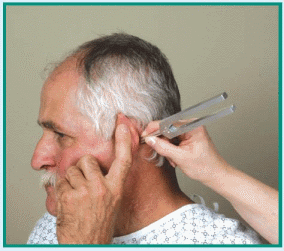
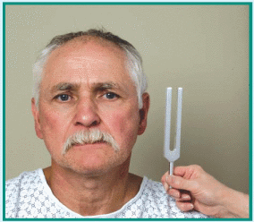
than age 50. Other physiologic causes of hearing loss include cerumen (earwax) impaction; barotitis media (unequal pressure on the eardrum) associated with descent in an airplane or elevator, diving, or close proximity to an explosion; and chronic exposure to noise over 90 decibels, which can occur on the job, with certain hobbies, or from listening to live or recorded music.


HISTORY AND PHYSICAL EXAMINATION

If the patient reports hearing loss, ask him to describe it fully. Is it unilateral or bilateral? Continuous or intermittent? Ask about a family history of hearing loss. Then obtain the patient’s medical history, noting chronic ear infections, ear surgery, and ear or head trauma. Has the patient recently had an upper respiratory tract infection? After taking a drug history, have the patient describe his occupation and work environment.

Next, explore associated signs and symptoms. Does the patient have ear pain? If so, is it unilateral or bilateral? Continuous or intermittent? Ask the patient if he has noticed discharge from one or both ears. If so, have him describe its color and consistency, and note when it began. Does he hear ringing, buzzing, hissing, or other noises in one or both ears? If so, are the noises constant or intermittent? Does he experience any dizziness? If so, when did he first notice it?

Begin the physical examination by inspecting the external ear for inflammation, boils, foreign bodies, and discharge. Then apply pressure to the tragus and mastoid to elicit tenderness. If you detect tenderness or external ear abnormalities, ask the physician whether an otoscopic examination should be done. (See Using an otoscope correctly, page 255.) During the otoscopic examination, note any color change, perforation, bulging, or retraction of the tympanic membrane, which normally looks like a shiny, pearl gray cone.

Next, evaluate the patient’s hearing acuity, using the ticking watch and whispered voice tests. Then perform the Weber and Rinne tests to obtain a preliminary evaluation of the type and degree of hearing loss. (See Differentiating conductive from sensorineural hearing loss, page 350.)


MEDICAL CAUSES

Acoustic neuroma. This eighth cranial nerve tumor causes unilateral, progressive, sensorineural hearing loss. The patient may also develop tinnitus, vertigo, and—with cranial nerve compression—facial paralysis.

Adenoid hypertrophy. Eustachian tube dysfunction gradually causes conductive hearing loss accompanied by intermittent ear discharge. The patient also tends to breathe through his mouth and may complain of a sensation of ear fullness.

Allergies. Conductive hearing loss may result when an allergy produces eustachian tube and middle ear congestion. Other features include ear pain or a feeling of fullness, nasal congestion, and conjunctivitis.

Aural polyps. If a polyp occludes the external auditory canal, partial hearing loss may occur. The polyp typically bleeds easily and is covered by a purulent discharge.

Cholesteatoma. Gradual hearing loss is characteristic in this disorder and may be accompanied by vertigo and, at times, facial paralysis. Examination reveals eardrum perforation, pearly white balls in the ear canal and, possibly, a discharge.

Cyst. Ear canal obstruction by a sebaceous or dermoid cyst causes progressive conductive hearing loss. On inspection, the cyst looks like a soft mass.

External ear canal tumor (malignant). Progressive conductive hearing loss is characteristic and is accompanied by deep, boring ear pain; a purulent discharge; and eventually facial paralysis. Examination may detect the granular, bleeding tumor.

Furuncle. Reversible conductive hearing loss may occur when one of these painful, hard nodules forms in the ear. The patient may report a sense of fullness in the ear and pain on palpation of the tragus or auricle. Rupture relieves the pain and produces a purulent, necrotic discharge.

Glomus jugulare tumor. Initially, this benign tumor causes mild, unilateral conductive hearing loss that becomes progressively more severe. The patient may report tinnitus that sounds like his heartbeat. Associated signs and symptoms include gradual congestion in the affected ear, throbbing or pulsating discomfort, bloody otorrhea, facial nerve paralysis, and vertigo. Although the tympanic membrane is normal, a reddened mass appears behind it.

Glomus tympanum tumor. This cancerous middle ear tumor causes slowly progressive hearing loss and throbbing or pulsating tinnitus.

It usually bleeds easily when manipulated. Late features include ear pain, dizziness, and total unilateral deafness.


Granuloma. A rare cause of conductive hearing loss, a granuloma may also produce fullness in the ear, deep-seated pain, and a bloody discharge.

Head trauma. Sudden conductive or sensorineural hearing loss may result from ossicle disruption, ear canal fracture, tympanic membrane perforation, or cochlear fracture associated with head trauma. Typically, the patient reports a headache and exhibits bleeding from his ear. Neurologic features vary and may include impaired vision and altered level of consciousness.

Herpes zoster oticus (Ramsay Hunt syndrome). This syndrome causes sudden severe, unilateral mixed hearing loss, which may be accompanied by vesicles in the external ear, tinnitus, vertigo, ear pain, malaise, and transient ipsilateral facial paralysis.

Hypothyroidism. This disorder may produce reversible sensorineural hearing loss. Other effects include bradycardia, weight gain despite anorexia, mental dullness, cold intolerance, facial edema, brittle hair, and dry skin that’s pale, cool, and doughy.

Ménière’s disease. Initially, this inner ear disorder produces intermittent, unilateral sensorineural hearing loss that involves only low tones. Later, hearing loss becomes constant and affects other tones. Associated signs and symptoms include intermittent severe vertigo, nausea and vomiting, a feeling of fullness in the ear, a roaring or hollow-seashell tinnitus, diaphoresis, and nystagmus.

Multiple sclerosis. Rarely, this disorder causes sensorineural hearing loss associated with myelin destruction of the central auditory pathways. The hearing loss may be sudden and unilateral or intermittent and bilateral. Among other characteristics are impaired vision, paresthesia, muscle weakness, gait ataxia, intention tremor, urinary disturbances, and emotional lability.

Myringitis. Rarely, acute infectious myringitis produces conductive hearing loss when fluid accumulates in the middle ear or a large bleb totally obstructs the ear canal. Small, reddened inflamed blebs may develop in the canal, on the tympanic membrane, or in the middle ear and may produce a bloody discharge if they rupture. Associated findings may include severe ear pain, mastoid tenderness, and fever.

Chronic granular myringitis produces gradual hearing loss accompanied by pruritus and a purulent discharge.

Nasopharyngeal cancer. This type of cancer causes mild unilateral conductive hearing loss when it compresses the eustachian tube. Bone conduction is normal, and inspection reveals a retracted tympanic membrane backed by fluid. When this tumor obstructs the nasal airway, the patient may exhibit nasal speech and a bloody nasal and postnasal discharge. Cranial nerve involvement produces other findings, such as diplopia and rectus muscle paralysis.

Osteoma. Commonly affecting women and swimmers, osteoma may cause sudden or intermittent conductive hearing loss. Typically, bony projections are visible in the ear canal, but the tympanic membrane appears normal.

Otitis externa. Conductive hearing loss resulting from debris in the ear canal characterizes both acute and malignant otitis externa. In acute otitis externa, ear canal inflammation produces pain, itching, and a foul-smelling, sticky yellow discharge. Severe tenderness is typically elicited by chewing, opening the mouth, and pressing on the tragus or mastoid. The patient may also develop a low-grade fever, regional lymphadenopathy, a headache on the affected side, and mild to moderate pain around the ear that may later intensify. Examination may reveal greenish white debris or edema in the canal.

In malignant otitis externa, debris is also visible in the canal. This life-threatening disorder, which most commonly occurs in diabetics, causes sensorineural hearing loss, pruritus, tinnitus, and severe ear pain.

Otitis media. This middle ear inflammation typically produces unilateral conductive hearing loss. In acute suppurative otitis media, the hearing loss develops gradually over a few hours and is usually accompanied by an upper respiratory tract infection with sore throat, cough, nasal discharge, and headache. Related signs and symptoms include dizziness, a sensation of fullness in the ear, intermittent or constant ear pain, fever, nausea, and vomiting. Rupture of the bulging, swollen tympanic membrane relieves the pain and produces a brief bloody and purulent discharge. Hearing returns after the infection subsides.

Hearing loss also develops gradually in patients with chronic otitis media. Assessment may reveal a perforated tympanic membrane, purulent ear drainage, earache, nausea, and vertigo.


Commonly associated with an upper respiratory tract infection or nasopharyngeal cancer, serous otitis media commonly produces a stuffy feeling in the ear and pain that worsens at night. Examination reveals a retracted—and perhaps discolored—tympanic membrane and possibly air bubbles behind the membrane.

Otosclerosis. In this hereditary disorder, unilateral conductive hearing loss usually begins when the patient is in his early twenties and may gradually progress to bilateral mixed hearing loss. The patient may report tinnitus and an ability to hear better in a noisy environment.

image Otosclerosis affects twice as many women as men and may worsen during pregnancy.

Skull fracture. Auditory nerve injury causes sudden unilateral sensorineural hearing loss. Accompanying signs and symptoms include ringing tinnitus, blood behind the tympanic membrane, scalp wounds, and other findings.

Syphilis. In tertiary syphilis, sensorineural hearing loss may develop suddenly or gradually and usually affects one ear more than the other. It’s usually accompanied by a gumma lesion—a chronic, superficial nodule or a deep, granulomatous lesion on the skin or mucous membranes. The lesion is solitary, asymmetrical, painless, and indurated. The patient may also exhibit signs of liver, respiratory, cardiovascular, or neurologic dysfunction.

Temporal arteritis. This disorder may produce unilateral sensorineural hearing loss accompanied by throbbing unilateral facial pain, pain behind the eye, temporal or frontotemporal headache, and occasionally vision loss. The hearing loss is usually preceded by a prodrome of malaise, anorexia, weight loss, weakness, and myalgia that lasts for several days. Examination may reveal a nodular, swollen temporal artery. Low-grade fever, confusion, and disorientation may also occur.

Temporal bone fracture. This fracture can cause sudden unilateral sensorineural hearing loss accompanied by hissing tinnitus. The tympanic membrane may be perforated, depending on the fracture’s location. Loss of consciousness, Battle’s sign, and facial paralysis may also occur.

Tuberculosis. This pulmonary infection may spread to the ear, resulting in eardrum perforation, mild conductive hearing loss, and cervical lymphadenopathy.

Tympanic membrane perforation. Commonly caused by trauma from sharp objects or rapid pressure changes, perforation of the tympanic membrane causes abrupt hearing loss along with ear pain, tinnitus, vertigo, and a sensation of fullness in the ear.

Wegener’s granulomatosis. Conductive hearing loss develops slowly in this rare necrotizing, granulomatous vasculitis. This multisystem disorder may also cause cough, pleuritic chest pain, epistaxis, hemorrhagic skin lesions, oliguria, and nasal discharge.


OTHER CAUSES

Drugs. Ototoxic drugs typically produce ringing or buzzing tinnitus and a feeling of fullness in the ear. Chloroquine, cisplatin, vancomycin, and aminoglycosides (especially neomycin, kanamycin, and amikacin) may cause irreversible hearing loss. Loop diuretics, such as furosemide, ethacrynic acid, and bumetanide, usually produce a brief, reversible hearing loss. Quinine, quinidine, and high doses of erythromycin or salicylates (such as aspirin) may also cause reversible hearing loss.

Radiation therapy. Irradiation of the middle ear, thyroid, face, skull, or nasopharynx may cause eustachian tube dysfunction, resulting in hearing loss.

Surgery. Myringotomy, myringoplasty, simple or radical mastoidectomy, or fenestrations may cause scarring that interferes with hearing.


SPECIAL CONSIDERATIONS

When talking with the patient, remember to face him and speak slowly. Don’t shout at the patient or smoke, eat, or chew gum when talking.

Prepare the patient for audiometry and auditory evoked-response testing. After testing, the patient may require a hearing aid or cochlear implant to improve his hearing.


PEDIATRIC POINTERS

About 3,000 profoundly deaf infants are born in the United States each year. In about half of these infants, hereditary disorders (such as Paget’s disease and Alport’s, Hurler’s, and Klippel-Feil syndromes) cause the typically sensorineural hearing loss. Nonhereditary disorders associated with congenital sensorineural hearing loss include albinism, onychodystrophy, cochlear dysplasia, and Pendred’s, Usher’s, Waardenburg’s, and Jervell and Lange-Nielsen syndromes. Sensorineural hearing loss may also result from maternal use of ototoxic drugs, birth trauma, and anoxia during or after birth.


Mumps is the most common cause of unilateral sensorineural hearing loss in children. Other causes are meningitis, measles, influenza, and acute febrile illness.

Congenital conductive hearing loss may be caused by atresia, ossicle malformation, and other abnormalities. Serous otitis media commonly causes bilateral conductive hearing loss in children. Putting foreign objects in the ears can also cause conductive hearing loss.

Hearing disorders in children may lead to speech, language, and learning problems. Early identification and treatment of hearing loss is thus crucial to avoid incorrectly labeling the child as mentally retarded, brain damaged, or a slow learner.

When assessing an infant or a young child for hearing loss, remember that you can’t use a tuning fork. Instead, test the startle reflex in infants younger than age 6 months, or have an audiologist test brain stem evoked response in neonates, infants, and young children. Also, obtain a gestational, perinatal, and family history from the parents.


GERIATRIC POINTERS

In older patients, presbycusis may be aggravated by exposure to noise as well as other factors.


PATIENT COUNSELING

Instruct the patient to avoid exposure to loud noise and to use ear protection to arrest hearing loss. If the patient has an upper respiratory tract infection, tell him to avoid flying and driving.


Heat intolerance

Heat intolerance refers to the inability to withstand high temperatures or to maintain a comfortable body temperature. This symptom produces a continuous feeling of being overheated and, at times, profuse diaphoresis. It usually develops gradually and is chronic.

Most cases of heat intolerance result from thyrotoxicosis. In this disorder, excess thyroid hormone stimulates peripheral tissues, increasing basal metabolism and producing excess heat. Although rare, hypothalamic disease may also cause intolerance to heat and cold.


HISTORY AND PHYSICAL EXAMINATION

Ask the patient when he first noticed his heat intolerance. Did he gradually use fewer blankets at night? Does he have to turn up the air conditioning to keep cool? Is it hard for him to adjust to warm weather? Does he sweat a lot in a hot environment? Find out if his appetite or weight has changed. Also, ask about unusual nervousness or other personality changes. Then take a drug history, especially noting use of amphetamines or amphetamine-like drugs. Ask the patient if he takes a thyroid drug. If so, what is the daily dosage and when did he last take it?

As you begin the examination, notice how much clothing the patient is wearing. After taking vital signs, inspect the patient’s skin for flushing and diaphoresis. Also, note tremors and lid lag.


MEDICAL CAUSES

Hypothalamic disease. In this rare disease, body temperature fluctuates dramatically, causing alternating heat and cold intolerance. Related features include amenorrhea, disturbed sleep patterns, increased thirst and urination, increased appetite with weight gain, impaired visual acuity, headache, and personality changes, such as bursts of rage or laughter. Common causes of hypothalamic disease are pituitary adenoma and hypothalamic and pineal tumors.

Thyrotoxicosis. A classic symptom of thyrotoxicosis, heat intolerance may be accompanied by an enlarged thyroid gland, nervousness, weight loss despite increased appetite, diaphoresis, diarrhea, tremor, and palpitations. Although exophthalmos is characteristic, many patients don’t display this sign. Associated findings may affect virtually every body system. Some common findings include irritability, difficulty concentrating, mood swings, insomnia, muscle weakness, fatigue, lid lag, tachycardia, full and bounding pulse, widened pulse pressure, dyspnea, amenorrhea, and gynecomastia. Typically, the patient’s skin is warm and flushed; premature graying and alopecia occur in both sexes.


OTHER CAUSES

Drugs. Amphetamines, amphetamine-like appetite suppressants, and excessive doses of thyroid hormone may cause heat intolerance. Anticholinergics may interfere with sweating, resulting in heat intolerance.


SPECIAL CONSIDERATIONS

Adjust room temperature to make the patient comfortable. If the patient is diaphoretic,
change his clothing and bed linens as necessary, and encourage him to drink lots of fluids.


PEDIATRIC POINTERS

Rarely, maternal thyrotoxicosis may be passed to the neonate, resulting in heat intolerance. More commonly, acquired thyrotoxicosis appears between ages 12 and 14, although this too is infrequent. Dehydration may also make a child sensitive to heat.


Heberden’s nodes

Heberden’s nodes are painless, irregular, cartilaginous or bony enlargements of the distal interphalangeal joints of the fingers. They reflect degeneration of articular cartilage, which irritates the bone and stimulates osteoblasts, causing bony enlargement. Approximately 2 to 3 mm in diameter, Heberden’s nodes develop on one or both sides of the dorsal midline. The dominant hand usually has larger nodes, which affect one or more fingers but not the thumb. (See Recognizing Heberden’s nodes.)

Osteoarthritis is the most common cause of Heberden’s nodes; in fact, more than one-half of all osteoarthritic patients have these nodes. Less commonly, repeated fingertip trauma may lead to node formation in only one joint (“baseball finger”). Because Heberden’s nodes aren’t associated with joint pain or loss of function, they aren’t a primary indicator of osteoarthritis; however, they’re a helpful adjunct to diagnosis.


HISTORY AND PHYSICAL EXAMINATION

Begin by asking the patient if anyone else in his family has had Heberden’s nodes or osteoarthritis. Are the patient’s joints stiff? Does stiffness disappear with movement? Ask him which hand is dominant. Also ask about repeated fingertip trauma associated with his job or sports.

Carefully palpate the nodes, noting any signs of inflammation, such as redness and tenderness. Then determine range of motion (ROM) in the fingers of each hand. As you do so, listen and feel for crepitation.


MEDICAL CAUSES

Osteoarthritis. This disorder commonly causes Heberden’s nodes and may also cause nodes in the proximal interphalangeal joints (Bouchard’s nodes). Its chief symptom, though, is joint pain that’s aggravated by movement or weight bearing. Joints may also be tender and display restricted ROM. Typically, joint stiffness is triggered by disuse and relieved by brief exercise. Stiffness may be accompanied by bony enlargement and crepitus.


SPECIAL CONSIDERATIONS

Remind the patient to take an anti-inflammatory drug and to exercise regularly. Encourage him to avoid joint strain, for example, by maintaining a healthy body weight.


PEDIATRIC POINTERS

Because children don’t suffer from osteoarthritis, they don’t develop Heberden’s nodes.


Hematemesis

Hematemesis, the vomiting of blood, usually indicates GI bleeding above the ligament of Treitz, which suspends the duodenum at its junction with the jejunum. Bright red or blood-streaked vomitus indicates fresh or recent bleeding. Dark red, brown, or black vomitus (the color and consistency of coffee grounds) indicates that blood has been retained in the stomach and partially digested.

Although hematemesis usually results from a GI disorder, it may stem from a coagulation disorder or from a treatment that irritates the GI tract. Swallowed blood from epistaxis or oropharyngeal erosion may also cause bloody vomitus. Hematemesis may be precipitated by straining, emotional stress, and the use of
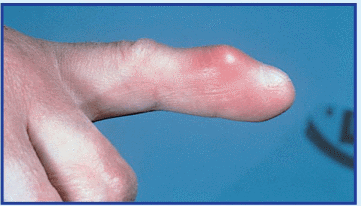
anti-inflammatory drugs or alcohol. In a patient with esophageal varices, hematemesis may be due to trauma from swallowing hard or partially chewed food. (See Rare causes of hematemesis.)

Hematemesis is always an important sign, but its severity depends on the amount, source, and intensity of the bleeding. Massive hematemesis (vomiting of 500 to 1,000 ml of blood) may be life-threatening.

image 
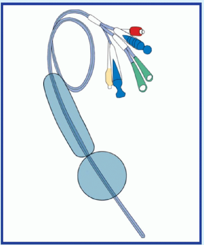
If the patient has massive hematemesis, check his vital signs. If you detect signs of shock—such as tachypnea, hypotension, and tachycardia—place the patient in a supine position, and elevate his feet 20 to 30 degrees. Start a large-bore I.V. catheter for emergency fluid replacement. Also, obtain a blood sample for typing and crossmatching, hemoglobin level, and hematocrit, and administer oxygen. Emergency endoscopy may be necessary to locate the source of bleeding. Prepare to insert a nasogastric (NG) tube for suction or iced lavage. A Sengstaken-Blakemore tube may be used to compress esophageal varices. (See Managing hematemesis with intubation, page 356.)
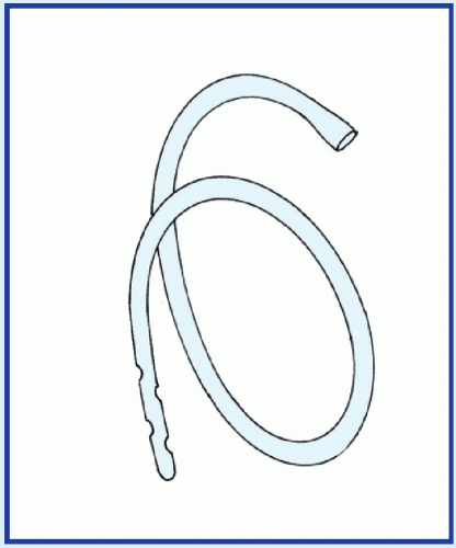
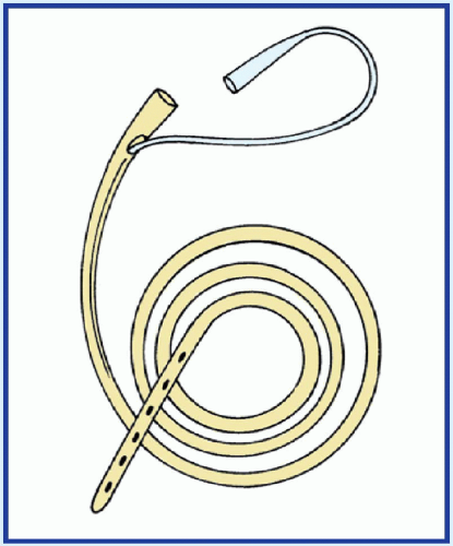

HISTORY AND PHYSICAL EXAMINATION

If hematemesis isn’t immediately life-threatening, begin with a thorough history. First, have the patient describe the amount, color, and consistency of the vomitus. When did he first notice this sign? Has he ever had hematemesis before? Find out if he also has bloody or black tarry stools. Note whether hematemesis is usually preceded by nausea, flatulence, diarrhea, or weakness. Has he recently had bouts of retching with or without vomiting?

Next, ask about a history of ulcers or of liver or coagulation disorders. Find out how much alcohol the patient drinks, if any. Does he regularly take aspirin or another nonsteroidal anti-inflammatory drug (NSAID), such as phenylbutazone or indomethacin? These drugs may cause erosive gastritis or ulcers.

Begin the physical examination by checking for orthostatic hypotension, an early warning sign of hypovolemia. Take blood pressure and pulse with the patient in the supine, sitting, and standing positions. A decrease of 10 mm Hg or more in systolic pressure or an increase of 10 beats/minute or more in pulse rate indicates volume depletion. After obtaining other vital signs, inspect the mucous membranes, nasopharynx, and skin for any signs of bleeding or other abnormalities. Finally, palpate the abdomen for tenderness, pain, or masses. Note lymphadenopathy.


MEDICAL CAUSES

Achalasia. Hematemesis is a rare effect of this disorder, which usually causes passive regurgitation and painless, progressive dysphagia. Regurgitation of undigested food may cause hoarseness, coughing, aspiration, and recurrent pulmonary infections.

Anthrax, GI. GI anthrax is caused by eating meat contaminated with the gram-positive, spore-forming bacterium Bacillus anthracis. Initial signs and symptoms of anorexia, nausea, vomiting, and fever may progress to hematemesis, abdominal pain, and severe bloody diarrhea.

Coagulation disorders. Any disorder that disrupts normal clotting, such as thrombocytopenia or hemophilia, may result in GI bleeding and moderate to severe hematemesis. Bleeding may occur in other body systems as well, resulting in such signs as epistaxis and ecchymosis. Associated effects depend on the specific coagulation disorder.

Esophageal cancer. A late sign of this cancer, hematemesis may be accompanied by steady chest pain that radiates to the back. Other features include substernal fullness, severe dysphagia, nausea, vomiting with nocturnal regurgitation and aspiration, hemoptysis, fever, hiccups, sore throat, melena, and halitosis.


Esophageal injury by caustic substances. Ingestion of corrosive acids or alkalies produces esophageal injury associated with grossly bloody or coffee-ground vomitus. Hematemesis is accompanied by epigastric and anterior or retrosternal chest pain that’s intensified by swallowing. With ingestion of alkaline agents, the oral and pharyngeal mucosa may produce a soapy white film. The mucosa becomes brown and edematous with time. Dysphagia, marked salivation, and fever may develop in 3 to 4 weeks and worsen as strictures form.

Esophageal rupture. The severity of hematemesis depends on the cause of the rupture. When an instrument damages the esophagus, hematemesis is usually slight. However, rupture due to Boerhaave’s syndrome (increased esophageal pressure from vomiting or retching) or other esophageal disorders typically causes more severe hematemesis. This life-threatening disorder may also produce severe retrosternal, epigastric, neck, or scapular pain accompanied by chest and neck edema. Examination reveals subcutaneous crepitation in the chest wall,
supraclavicular fossa, and neck. The patient may also show signs of respiratory distress, such as dyspnea and cyanosis.

Esophageal varices (ruptured). Life-threatening rupture of esophageal varices may produce coffee-ground or massive bright red vomitus. Signs of shock, such as hypotension and tachycardia, may follow or even precede hematemesis if the stomach fills with blood before vomiting occurs. Other symptoms may include abdominal distention and melena or painless hematochezia (ranging from slight oozing to massive rectal hemorrhage).

Gastric cancer. Painless bright red or dark brown vomitus is a late sign of this uncommon cancer, which usually begins insidiously with upper abdominal discomfort. The patient then develops anorexia, mild nausea, and chronic dyspepsia that’s unrelieved by antacids and exacerbated by food. Later symptoms may include fatigue, weakness, weight loss, feelings of fullness, melena, altered bowel habits, and signs of malnutrition, such as muscle wasting and dry skin.

Gastritis (acute). Hematemesis and melena are the most common signs of acute gastritis. They may even be the only signs, although mild epigastric discomfort, nausea, fever, and malaise may also occur. Massive blood loss precipitates signs of shock. Typically, the patient has a history of alcohol abuse or has used aspirin or another NSAID. Gastritis may also occur secondary to Helicobacter pylori infection.

Gastroesophageal reflux disease. Although rare in this disorder, hematemesis may produce significant blood loss. It’s accompanied by pyrosis, flatulence, dyspepsia, and postural regurgitation that can be aggravated by lying down or stooping over. Related effects include dysphagia, retrosternal angina-like chest pain, weight loss, halitosis, and signs of aspiration, such as dyspnea and recurrent pulmonary infections.

Leiomyoma. This benign tumor occasionally involves the GI tract, eroding the mucosa or vascular supply to produce hematemesis. Other features vary with the tumor’s size and location. For example, esophageal involvement may cause dysphagia and weight loss.

Mallory-Weiss syndrome. Characterized by a mucosal tear of the mucous membrane at the junction of the esophagus and the stomach, this syndrome may produce hematemesis and melena. It’s commonly triggered by severe vomiting, retching, or straining (as from coughing), usually in alcoholics or in people whose pylorus is obstructed. Severe bleeding may precipitate signs of shock, such as tachycardia, hypotension, dyspnea, and cool, clammy skin.

Peptic ulcer. Hematemesis may occur when a peptic ulcer penetrates an artery, vein, or highly vascular tissue. Massive—and possibly life-threatening—hematemesis is typical when an artery is penetrated. Other features include melena or hematochezia, chills, fever, and signs and symptoms of shock and dehydration, such as tachycardia, hypotension, poor skin turgor, and thirst. Most patients have a history of nausea, vomiting, epigastric tenderness, and epigastric pain that’s relieved by foods or antacids. Some may also have a history of habitual use of tobacco, alcohol, or NSAIDs.


OTHER CAUSES

Treatments. Traumatic NG or endotracheal intubation may cause hematemesis associated with swallowed blood. Nose or throat surgery may also cause this sign in the same way.


SPECIAL CONSIDERATIONS

Closely monitor the patient’s vital signs, and watch for signs of shock. Check the patient’s stools regularly for occult blood, and keep accurate intake and output records. Place the patient on bed rest in a low or semi-Fowler’s position to prevent aspiration of vomitus. Keep suctioning equipment nearby, and use it as needed. Provide frequent oral hygiene and emotional support—the sight of bloody vomitus can be very frightening. Administer a histamine-2 blocker I.V.; vasopressin may be required for ruptured esophageal varices. As the bleeding tapers off, monitor the pH of gastric contents, and give hourly doses of antacids by NG tube as necessary.


PEDIATRIC POINTERS

Hematemesis is much less common in children than in adults and may be related to foreignbody ingestion. Occasionally, neonates develop hematemesis after swallowing maternal blood during delivery or breast-feeding from a cracked nipple. Hemorrhagic disease of the neonate and esophageal erosion may also cause hematemesis in infants; such cases require immediate fluid replacement.


GERIATRIC POINTERS

In elderly patients, hematemesis may be caused by a vascular anomaly, an aortoenteric fistula, or upper GI cancer. In addition, chronic
obstructive pulmonary disease, chronic hepatic or renal failure, and chronic NSAID use all predispose elderly people to hemorrhage secondary to coexisting ulcerative disorders.


PATIENT COUNSELING

Explain diagnostic tests, such as endoscopy, barium swallow, and variceal banding. Explain laboratory tests, such as serum electrolyte levels, complete blood count, prothrombin time, partial thromboplastin time, and international normalized ratio.


Hematochezia

[Rectal bleeding]

The passage of bloody stools, also known as hematochezia, usually indicates—and may be the first sign of—GI bleeding below the ligament of Treitz. However, this sign—usually preceded by hematemesis—may also accompany rapid hemorrhage of 1 L or more from the upper GI tract.

Hematochezia ranges from formed, bloodstreaked stools to liquid, bloody stools that may be bright red, dark mahogany, or maroon in color. This sign usually develops abruptly and is heralded by abdominal pain.

Although hematochezia is commonly associated with GI disorders, it may also result from a coagulation disorder, exposure to toxins, or certain diagnostic tests. Always a significant sign, hematochezia may precipitate life-threatening hypovolemia.

image If the patient has severe hematochezia, check his vital signs. If you detect signs of shock, such as hypotension and tachycardia, place the patient in a supine position and elevate his feet 20 to 30 degrees. Prepare to administer oxygen, and start a large-bore I.V. catheter for emergency fluid replacement. Next, obtain a blood sample for typing and crossmatching, hemoglobin level, and hematocrit. Insert a nasogastric tube. Iced lavage may be indicated to control bleeding. Endoscopy may be necessary to detect the source of the bleeding.


HISTORY AND PHYSICAL EXAMINATION

If the hematochezia isn’t immediately life-threatening, ask the patient to fully describe the amount, color, and consistency of his bloody stools. (If possible, also inspect and characterize the stools yourself.) How long have the stools been bloody? Do they always look the same, or does the amount of blood seem to vary? Ask about associated signs and symptoms.

Next, explore the patient’s medical history, focusing on GI and coagulation disorders. Ask about the use of GI irritants, such as alcohol, aspirin, and other nonsteroidal anti-inflammatory drugs.

Begin the physical examination by checking for orthostatic hypotension, an early sign of shock. Take the patient’s blood pressure and pulse while he’s lying down, sitting, and standing. If systolic pressure decreases by 10 mm Hg or more, or pulse rate increases by 10 beats/minute or more when he changes position, suspect volume depletion and impending shock.

Examine the skin for petechiae or spider angiomas. Palpate the abdomen for tenderness, pain, or masses. Also, note lymphadenopathy. Finally, a digital rectal examination must be done to rule out rectal masses or hemorrhoids.


MEDICAL CAUSES

Amyloidosis. Hematochezia occasionally occurs when this disorder affects the GI tract. Massive, rapid hematochezia may precipitate signs of shock, such as hypotension and tachycardia. Associated signs and symptoms include hypoactive or absent bowel sounds, abdominal pain, malabsorption, diarrhea, and renal disease. The patient may also have a stiff, enlarged tongue, resulting in dysarthria.

Anal fissure. Slight hematochezia characterizes this disorder; blood may streak the stools or appear on toilet tissue. Accompanying hematochezia is severe rectal pain that may make the patient reluctant to defecate, thereby causing constipation.

Angiodysplastic lesions. Most common in elderly patients, these arteriovenous lesions of the ascending colon typically cause chronic, bright red rectal bleeding. Occasionally, they may result in life-threatening blood loss and signs of shock, such as tachycardia and hypotension.

Anorectal fistula. Blood, pus, mucus, and occasionally stools may drain from this type of fistula. Other effects include rectal pain and pruritus.

Coagulation disorders. Patients with a coagulation disorder (such as thrombocytopenia or disseminated intravascular coagulation) may experience GI bleeding marked by moderate to severe hematochezia. Bleeding may also occur
in other body systems, producing such signs as epistaxis and purpura. Associated findings vary with the specific coagulation disorder.

Colitis. Ischemic colitis commonly causes bloody diarrhea, especially in elderly patients. Rectal bleeding may be slight or massive and is usually accompanied by severe, cramping lower abdominal pain and hypotension. Other effects include abdominal tenderness, distention, and absent bowel sounds. Severe colitis may cause life-threatening hypovolemic shock and peritonitis.

Ulcerative colitis typically causes bloody diarrhea that may also contain mucus. Blood loss may be slight or massive and is preceded by mild to severe abdominal cramps. Associated signs and symptoms include fever, tenesmus, anorexia, nausea, vomiting, hyperactive bowel sounds and, occasionally, tachycardia. Weight loss and weakness occur late.

Colon cancer. Bright red rectal bleeding with or without pain is a telling sign, especially in cancer of the left colon. This type of tumor usually causes early signs of obstruction, such as rectal pressure, bleeding, and intermittent fullness or cramping. As the disease progresses, the patient also develops obstipation, diarrhea or ribbon-shaped stools, and pain that’s typically relieved by passage of stools or flatus. Stools are grossly bloody.

Cancer of the right colon may initially cause melena and abdominal aching, pressure, and dull cramps. As the disease progresses, the patient may also experience diarrhea, anorexia, weight loss, anemia, weakness and fatigue, vomiting, an abdominal mass, and signs of obstruction, such as abdominal distention and abnormal bowel sounds.

Colorectal polyps. These polyps are the most common cause of intermittent hematochezia in adults younger than age 60, but they don’t always produce symptoms. When located high in the colon, polyps may cause bloodstreaked stools that yield a positive response when tested with guaiac. If the polyps are located closer to the rectum, they may bleed freely.

Crohn’s disease. Hematochezia is not a common sign of this disorder unless the perineum is involved. If rectal bleeding does occur, it’s likely to be massive. The chief clinical features of Crohn’s disease include fever, abdominal distention and pain with guarding, diarrhea, hyperactive bowel sounds, anorexia, nausea, and fatigue. Palpation may reveal a mass in the colon.

Diverticulitis. Most common in elderly patients, this disorder can suddenly cause mild to moderate rectal bleeding after the patient feels the urge to defecate. The bleeding may end abruptly or may progress to life-threatening blood loss with signs of shock. Associated signs and symptoms may include left-lower-quadrant pain that’s relieved by defecation, alternating episodes of constipation and diarrhea, anorexia, nausea and vomiting, rebound tenderness, and a distended tympanic abdomen.

Dysentery. Bloody diarrhea is common in infection with Shigella, Amoeba, and Campylobacter, but rare with Salmonella. Abdominal pain or cramps, tenesmus, fever, and nausea may also occur.

Esophageal varices (ruptured). In this life-threatening disorder, hematochezia may range from slight rectal oozing to grossly bloody stools and may be accompanied by mild to severe hematemesis or melena. Signs of shock, such as tachycardia and hypotension, may follow or occasionally precede overt signs of bleeding. Typically, the patient has a history of chronic liver disease.

Food poisoning (staphylococcal). The patient may have bloody diarrhea 1 to 6 hours after ingesting food toxins. Accompanying signs and symptoms, which last a few hours, include severe, cramping abdominal pain, nausea and vomiting, and prostration.

Hemorrhoids. Hematochezia may accompany external hemorrhoids, which typically cause painful defecation, resulting in constipation. Less painful internal hemorrhoids usually produce more chronic bleeding with bowel movements, which may eventually lead to signs of anemia, such as weakness and fatigue.

Leptospirosis. The severe form of this infection—Weil’s syndrome—produces hematochezia or melena along with other signs of bleeding, such as epistaxis and hemoptysis. The bleeding is typically preceded by a sudden frontal headache, severe thigh and lumbar myalgia, cutaneous hyperesthesia, and conjunctival suffusion. Bleeding is followed by chills, a rapidly rising fever, and perhaps nausea and vomiting. Fever, headache, and myalgia usually intensify and persist for weeks. Other findings may include right-upper-quadrant tenderness, hepatomegaly, and jaundice.

Peptic ulcer. Upper GI bleeding is a common complication in this disorder. The patient may display hematochezia, hematemesis, or melena, depending on the intensity and amount of
bleeding. If the peptic ulcer penetrates an artery or vein, massive bleeding may precipitate signs of shock, such as hypotension and tachycardia. Other findings may include chills, fever, nausea and vomiting, and signs of dehydration, such as dry mucous membranes, poor skin turgor, and thirst. Most patients have a history of epigastric pain that’s relieved by foods or antacids; some also have a history of habitual use of tobacco, alcohol, or nonsteroidal anti-inflammatory drugs.

Rectal melanoma (malignant). This rare form of rectal cancer typically causes recurrent rectal bleeding that arises from a painless, asymptomatic mass.

Small-intestine cancer. This disorder occasionally produces slight hematochezia or bloodstreaked stools. Its characteristic features include colicky pain and postprandial vomiting. Other common signs and symptoms include anorexia, weight loss, and fever. Palpation may reveal abdominal masses.

Typhoid fever. About 10% of patients with typhoid fever develop hematochezia, which is occasionally massive. However, melena is more common. Both signs of bleeding occur late and may be accompanied by marked abdominal distention, diarrhea, significant weight loss, mental dullness, and profound fatigue. Earlier signs and symptoms are pathognomonic rose spots, headache, chills, fever, constipation, dry cough, conjunctivitis, and epistaxis.

Ulcerative proctitis. In this disorder, the patient typically has an intense urge to defecate but passes only bright red blood, pus, or mucus. Other common findings include acute constipation and tenesmus.


OTHER CAUSES

Heavy metal poisoning. Bloody diarrhea is accompanied by cramping abdominal pain, nausea, and vomiting. Other signs may include tachycardia, hypotension, seizures, paresthesia, depressed or absent deep tendon reflexes, and an altered level of consciousness.

Tests. Certain procedures, especially colonoscopy, polypectomy, and proctosigmoidoscopy, may cause rectal bleeding. Bowel perforation is rare.


SPECIAL CONSIDERATIONS

Place the patient on bed rest and check his vital signs frequently, watching for signs of shock, such as hypotension, tachycardia, weak pulse, and tachypnea. Monitor the patient’s intake and output hourly. Remember to provide emotional support because hematochezia may frighten the patient.

Prepare the patient for blood tests and GI procedures, such as endoscopy and GI X-rays. Visually examine the patient’s stools and test them for occult blood. If necessary, send a stool specimen to the laboratory to check for parasites.


PEDIATRIC POINTERS

Hematochezia is much less common in children than in adults. It may result from structural disorders, such as intussusception and Meckel’s diverticulum, and from inflammatory disorders, such as peptic ulcer disease and ulcerative colitis.

In children, ulcerative colitis typically produces chronic, rather than acute, signs and symptoms and may also cause slow growth and maturation related to malnutrition. Suspect sexual abuse in all cases of rectal bleeding in children.


GERIATRIC POINTERS

Because older people have an increased risk of colon cancer, hematochezia should be evaluated with colonoscopy after perirectal lesions have been ruled out as the cause of bleeding.


Hematuria

A cardinal sign of renal and urinary tract disorders, hematuria is the abnormal presence of blood in the urine. Strictly defined, it means three or more red blood cells (RBCs) per highpower microscopic field in the urine. Microscopic hematuria is confirmed by an occult blood test, whereas macroscopic hematuria is immediately visible. However, macroscopic hematuria must be distinguished from pseudohematuria. (See Confirming hematuria.) Macroscopic hematuria may be continuous or intermittent, is often accompanied by pain, and may be aggravated by prolonged standing or walking.

Hematuria may be classified by the stage of urination it predominantly affects. Bleeding at the start of urination—initial hematuria—usually indicates urethral pathology; bleeding at the end of urination—terminal hematuria—usually indicates pathology of the bladder neck, posterior urethra, or prostate; bleeding throughout urination —total hematuria—usually indicates pathology above the bladder neck.


Hematuria may result from one of two mechanisms: rupture or perforation of vessels in the renal system or urinary tract, or impaired glomerular filtration, which allows RBCs to seep into the urine. The color of the bloody urine provides a clue to the source of the bleeding. Generally, dark or brownish blood indicates renal or upper urinary tract bleeding, whereas bright red blood indicates lower urinary tract bleeding.

Although hematuria usually results from renal and urinary tract disorders, it may also result from certain GI, prostate, vaginal, or coagulation disorders or from the effects of certain drugs. Invasive therapy and diagnostic tests that involve manipulative instrumentation of the renal and urologic systems may also cause hematuria. Nonpathologic hematuria may result from fever and hypercatabolic states. Transient hematuria may follow strenuous exercise. (See Hematuria: Causes and associated findings, pages 362 to 365.)

Only gold members can continue reading. Log In or Register to continue

Stay updated, free articles. Join our Telegram channel

Aug 27, 2016 | Posted by in GENERAL & FAMILY MEDICINE | Comments Off on H

Full access? Get Clinical Tree

Get Clinical Tree app for offline access