FIGURE 15-1
(A) Lichen planus
(B) Lichen sclerosus et atrophicus
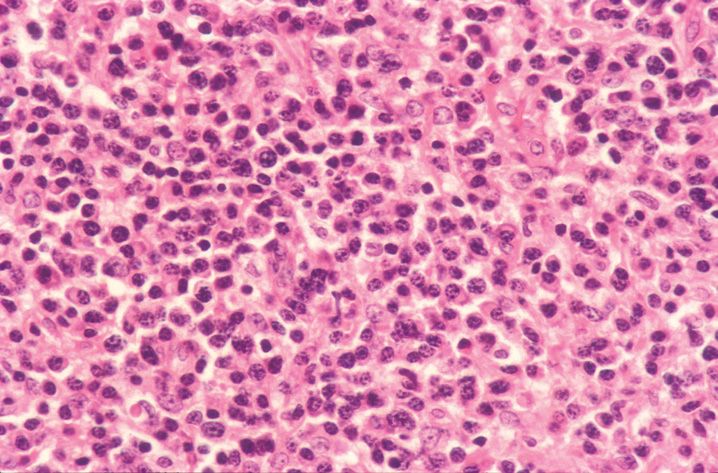
(C) Mycosis fungoides
(D) Plasmacytosis mucosae
(E) Syphilis
4. A 28-year-old woman has an indurated, painless vulvar ulcer. A biopsy is taken and is shown in Figure 15-2. Which stain would best highlight the causative organism?
(A) Fite stain
(B) Gomori methenamine silver stain
(C) Gram stain
(D) Herpes virus immunostain
(E) Warthin–Starry stain
5. A 42-year-old woman has a well-established diagnosis of psoriasis. Trauma (abrasion) to the vulva region results in the development of lesions. Trauma induced lesions are referred to as
(A) Auspitz sign
(B) Inverse psoriasis
(C) Koebner phenomenon
(D) Munroe sign
(E) Spongiform pustules of Kogoj
6. A 29-year-old woman presents with elevated, flat-topped, red-brown papules on the vulva. A diagnosis of condyloma lata is made. The causative organism is
(A) Candida albicans
(B) Chlamydia trachomatis
(C) Dermatophyte
(D) Herpes simplex
(E) Treponema pallidum
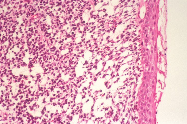
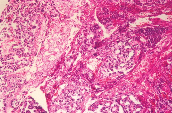
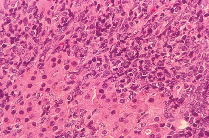
7. A biopsy is taken (shown in Figure 15-3) from an ulcerated vulvar lesion in a previously healthy 22-year-old woman. The most likely etiology is
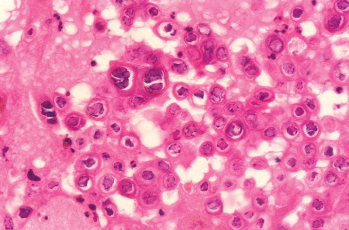
(A) Cytomegalovirus
(B) Epstein–Barr virus
(C) Herpes virus
(D) Rubella virus
(E) Varicella zoster virus
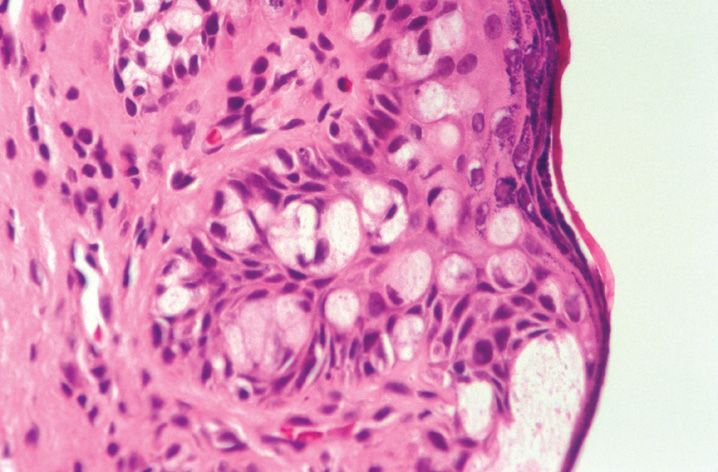
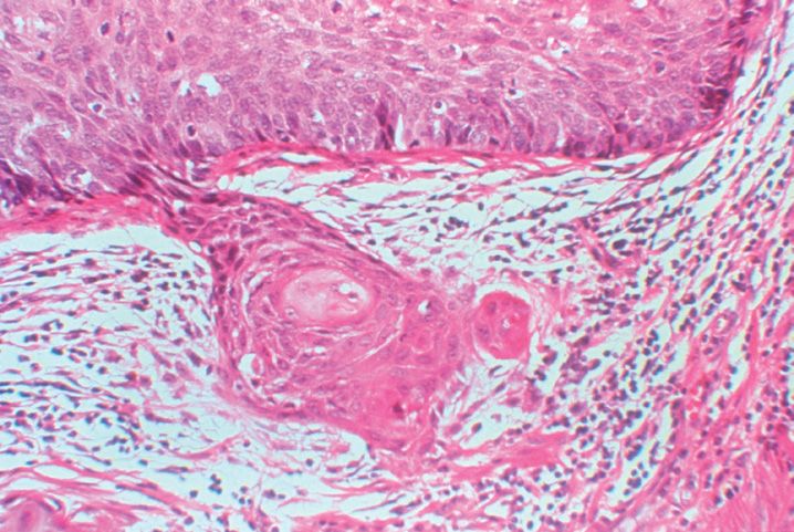
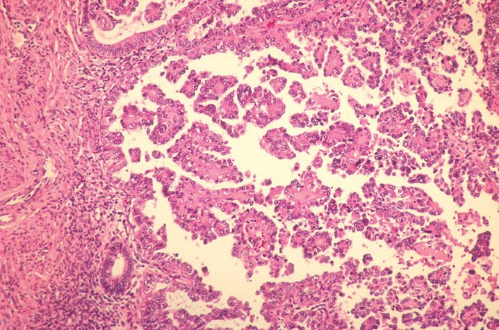
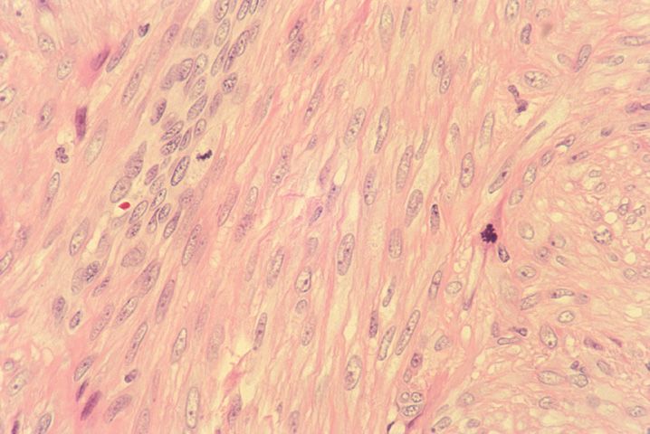
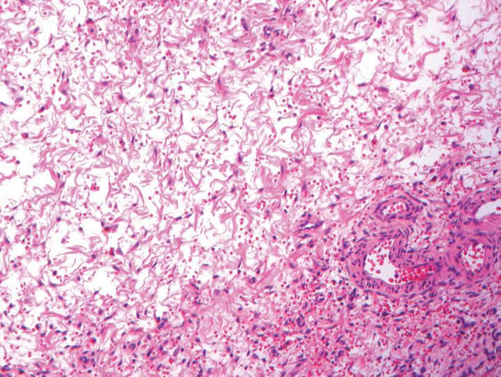
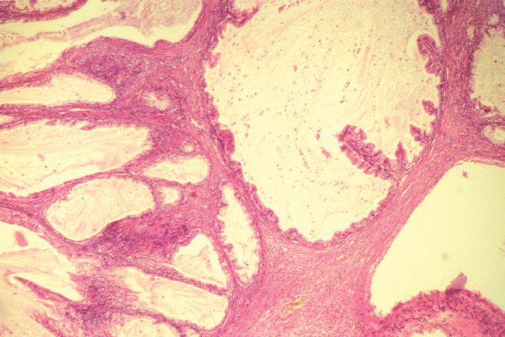
8. A 28-year-old pregnant woman presents with a 1 cm pedunculated mass on the vulva. A biopsy is taken and is shown in Figure 15-4. The most likely diagnosis is
(A) Aggressive angiomyxoma
(B) Botryoid rhabdomyosarcoma
(C) Condyloma acuminatum
(D) Fibroepithelial stromal polyp
(E) Malignant fibrosis histiocytoma
9. The stromal cells in the lesion in Question 8 is likely to stain with all of the following immunomarkers except
(A) CD34
(B) Desmin
(C) Estrogen receptor
(D) Progesterone receptor
(E) Vimentin
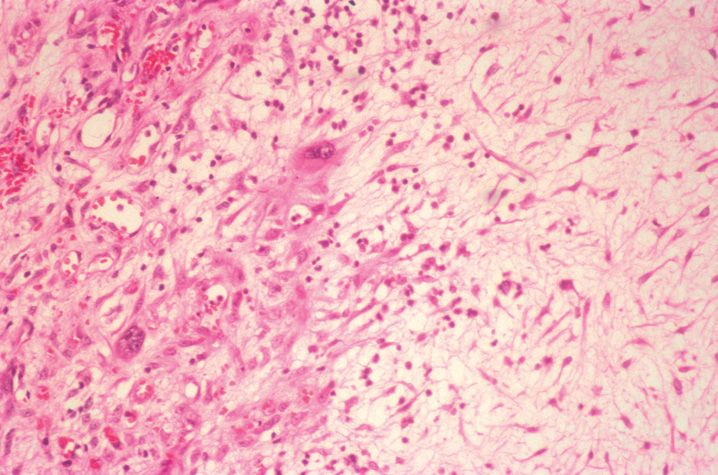
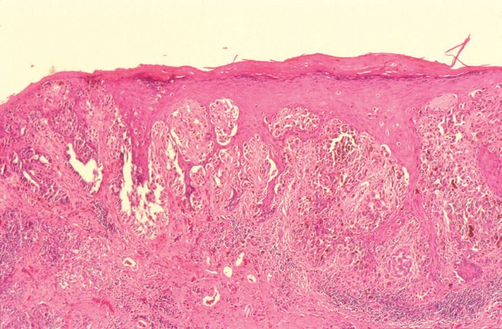
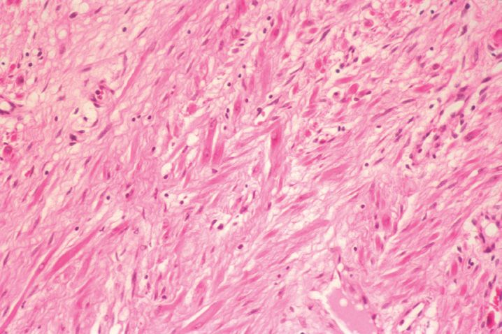
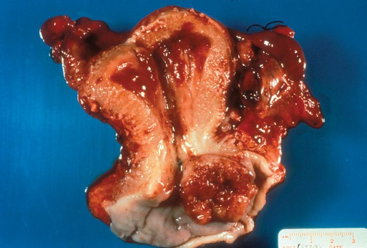
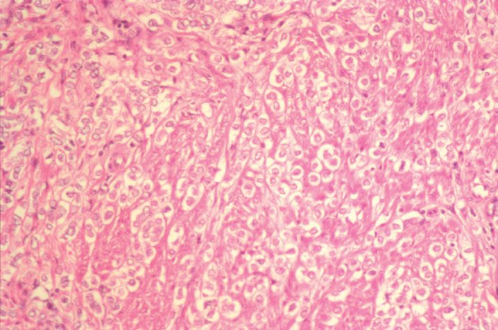
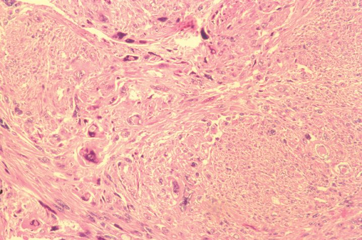
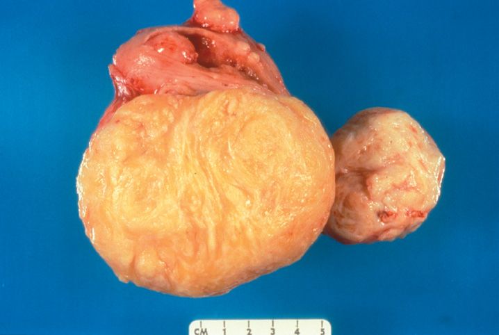
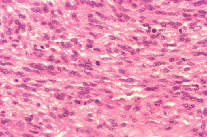
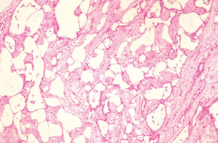
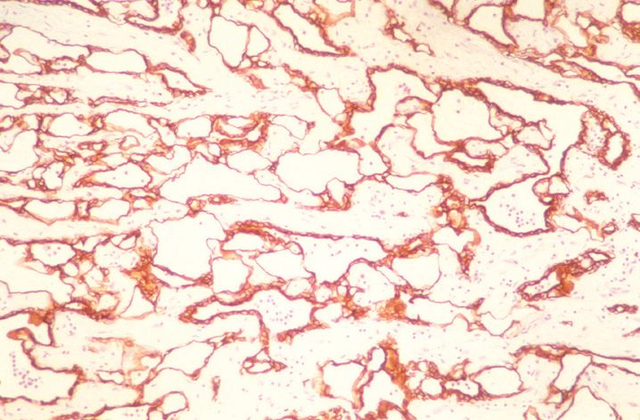
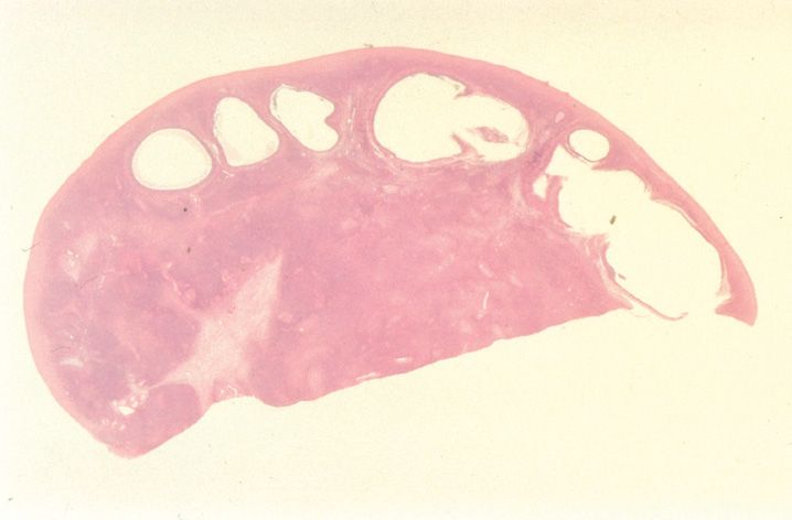
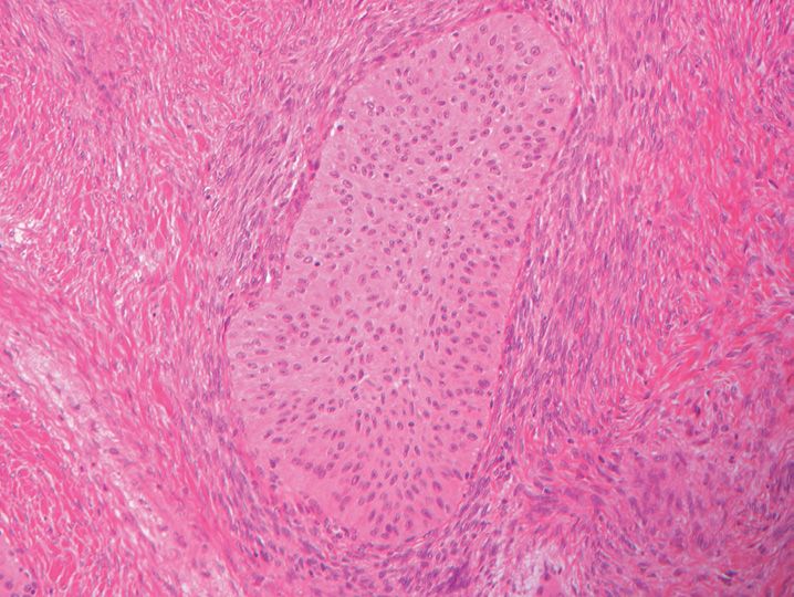
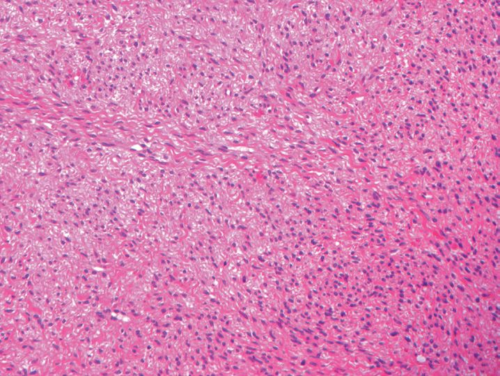
10. A 37-year-old woman presents with a perineal mass that measures 10 cm. She undergoes surgical resection of the mass, which is shown in Figure 15-5. The most likely diagnosis is
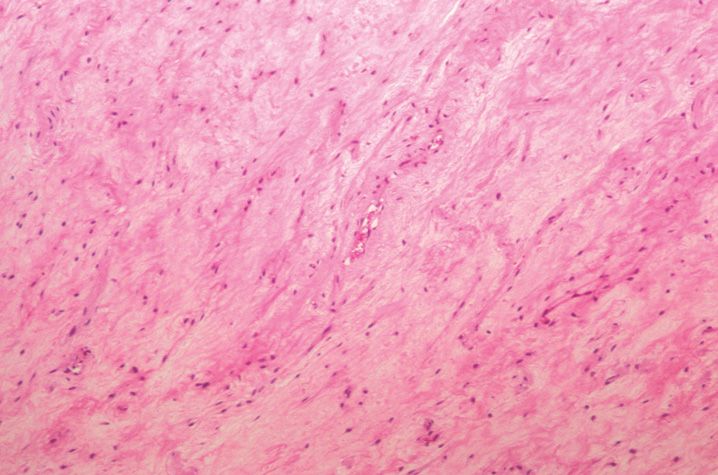
(A) Aggressive angiomyxoma
(B) Angiomyofibroblastoma
(C) Benign fibroepithelial polyp
(D) Cellular angiofibroma
(E) Liposarcoma
11. The most appropriate treatment for the lesion in Question 10 is
(A) Hormone therapy targeting estrogen/progesterone receptors
(B) None because it will spontaneously resolve
(C) Radiation alone
(D) Surgical excision with 1 cm margins
(E) Surgical excision with 1 cm margins and radiation
12. Which of the following is not true regarding cellular angiofibroma of the vulva?
(A) Mitotic figures are commonly seen.
(B) Most demonstrate CD34 immunoreactivity.
(C) They have minimal cytologic atypia.
(D) They typically arise in middle-aged women.
(E) Usually have an infiltrative border.
13. The lesion seen (Figure 15-6) in this 65-year-old woman is best classified as
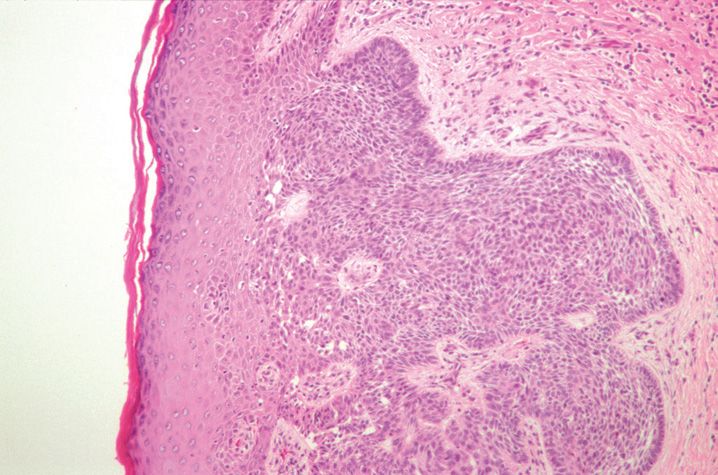
(A) Basal cell carcinoma
(B) Condyloma acuminatum
(C) Psoriasis
(D) Squamous cell carcinoma
(E) Verrucous carcinoma
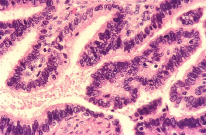
14. Which of the following immunomarkers would most likely be positive in this lesion from the vulva of a 41-year-old woman? (Figure 15-7)
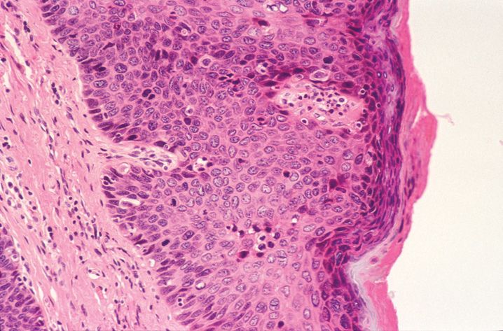
(A) CD19
(B) CD34
(C) CEA
(D) p16
(E) S-100 protein
15. A 39-year-old woman’s vulva lesion is stained with p53 antibody and suprabasilar staining of the surface epithelium is observed. This staining patten is seen with which pathology?
(A) Lichen sclerosus
(B) Lichen simplex chronicus
(C) Normal squamous mucosa
(D) Vulvar intraepithelial neoplasia
(E) Yeast infections
16. A 62-year-old woman has a vulvar carcinoma that is 1.3 cm in greatest dimension, confined to the vulva, and invades the stroma to a depth of 1.2 mm. The TNM stage of this tumor would be
(A) Tis
(B) T1a
(C) T1b
(D) T2
(E) T3
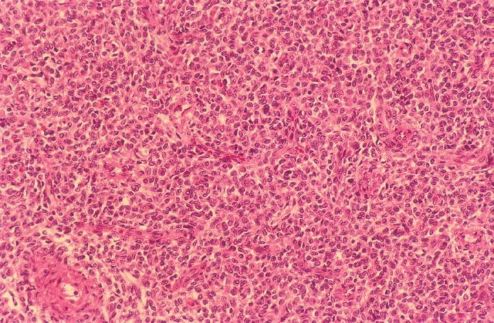
17. Which of the following is least predictive of outcome in the vulvar tumor seen (Figure 15-8) in a 75-year-old woman?
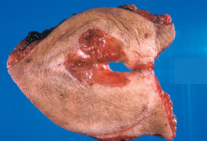
(A) Host inflammatory response
(B) Invasion depth
(C) Lymph node involvement
(D) Tumor stage
(E) Tumor size
18. The lesion shown in Figure 15-9 that arises in the vulva of a 54-year-old. It is best classified as which subtype of squamous cell carcinoma?
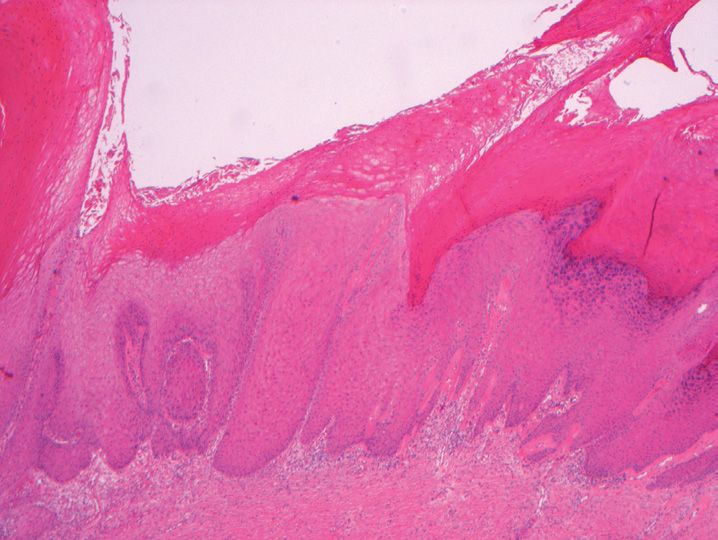
(A) Basaloid carcinoma
(B) Keratinizing squamous cell carcinoma
(C) Keratoacanthoma-like carcinoma
(D) Verrucous carcinoma
(E) Warty carcinoma
19. In differentiating between Merkel cell carcinoma and a basaloid squamous cell carcinoma of the vulva, all of the following are features typical of Merkel cell carcinoma except
(A) Cytokeratin 20 dot-like immunoreactivity
(B) Diffuse or trabecular growth pattern
(C) Frequent apoptosis
(D) Low mitotic index
(E) Tumor cells positive for neuron specific enolase
20. The least common site for the development of extramammary Paget disease is
(A) Anogenital region
(B) Axillae
(C) Ears
(D) Eyelids
(E) Sinuses
21. This vulvar lesion is encountered in a 62-year-old woman who presented with an eczematous, pruritic plaque. The lesion demonstrates the following immunohistochemical profile: cytokeratin 7 positive, cytokeratin 20 positive, GCDFP15 negative, uroplakin III positive, MUC2 negative. The most likely site of origin for the lesion is (Figure 15-10)
(A) Bladder
(B) Breast
(C) Cervix
(D) Colon
(E) Rectum
22. All of the following are true regarding this vulvar tumor except (Figure 15-11)
(A) Median survival is 3–4 years.
(B) Most commonly arises in the 3rd and 4th decades of life in this location.
(C) Second most common malignant vulvar neoplasm after squamous cell carcinoma.
(D) Stains positively with antibodies to MART1 and HMB-45.
(E) This is the most common location of this tumor in the female genital tract.
23. All of the following are common findings in the Vater syndrome except
(A) Anal atresia
(B) Deafness
(C) Renal malformations
(D) Single umbilical artery
(E) Vaginal atresia
24. A young woman presents with a thin watery vaginal discharge, a positive amine odor test, and a vaginal pH of 4.8. She is diagnosed with acute vaginitis. The most common cause of acute vaginitis is
(A) Bacterial vaginosis
(B) Candida infection
(C) Group B streptococcus
(D) Trauma
(E) Trichomonas vaginalis
25. A 34-year-old woman presents with pelvic pain and a malodorous vaginal discharge. She has an intrauterine device in place. Which of the following organisms is the most likely etiology of the vaginal abscess she is diagnosed with?
(A) Actinomyces
(B) Candida
(C) Group B streptococcus
(D) Herpes simplex type 2
(E) Trichomonas vaginalis
26. A 63-year-old woman presents with vaginal bleeding and a vaginal mass. A biopsy shows a prominent histocytic infiltrate with accompanying lymphocytes, plasma cells, and neutrophils. By electron microscopy, cytoplasmic structures are identified with an electron-lucent core, surrounded by radially oriented hydroxyapatite spicules. These findings are characteristic of which of the following?
(A) Emphysematous vaginitis
(B) Lichen planus
(C) Ligneous vaginitis
(D) Malakoplakia
(E) Toxic shock syndrome
27. A 26-year-old woman presents with a 2 cm antero-lateral wall vaginal cystic mass. The cyst is lined by cuboidal epithelium that does not stain with mucicarmine or PAS. The best diagnosis for this lesion would be which of the following?
(A) Cystic atrophy
(B) Epidermal inclusion cyst
(C) Gartner cyst
(D) Müllerian cyst
(E) Vaginal adenosis
28. All of the following are changes in the vagina associated with radiotherapy except
(A) Atrophic squamous epithelium
(B) Cells with cytoplasmic vacuolization
(C) Markedly increased mitotic figures
(D) Stromal hyalinization
(E) Vascular hyalinization
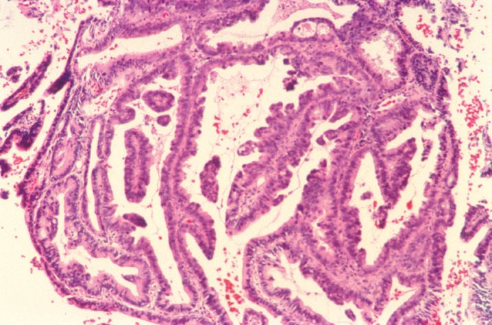
29. The lesion shown in Figure 15-12 arose in the vagina of a 28-year-old woman, who presented with vaginal bleeding and dyspareunia. The diagnosis is which of the following?
(A) Leiomyoma
(B) Müllerian papilloma
(C) Neurofibroma
(D) Rhabdomyoma
(E) Spindle cell epithelioma
30. All of the following are known risk factors for the development of vaginal squamous cell carcinoma except
(A) Alcohol
(B) Low socioeconomic status
(C) Pelvic radiation
(D) Smoking
(E) Trauma
31. A 46-year-old woman is diagnosed with vaginal squamous cell carcinoma. The tumor involves subvaginal tissue but does not extend beyond the pelvic wall. Which of the following is the correct International Federation of Gynecologists and Obstetrics (FIGO) stage?
(A) 0
(B) I
(C) II
(D) III
(E) IV
32. Which of the following vaginal lesions is most closely associated with a history of diethylstilbestrol (DES) exposure?
(A) Clear cell adenocarcinoma
(B) Endometrioid adenocarcinoma
(C) Mesonephric adenocarcinoma
(D) Mucinous adenocarcinoma
(E) Squamous cell carcinoma
33. This vaginal tumor shown in Figure 15-13 presented in a 4-year-old girl who presented with a mass protruding from the introitus. Which of the following immunomarkers would be expected to be positive in this lesion?
(A) Cytokeratin 7
(B) Cytokeratin 20
(C) Melan-A
(D) Myo-D1
(E) S-100 protein
34. A mass was removed from the vagina of a 2-year-old girl. The lesion stains with AFP but does not stain with CD30, PLAP, or cytokeratin 7. Which of the following is the correct diagnosis of this lesion? (Figure 15-14)
(B) Embryonal carcinoma
(C) Immature teratoma
(D) Metastatic dysgerminoma
(E) Yolk sac tumor
35. Tumors from which of the following sites are most likely to metastasize to vagina?
(A) Bladder
(B) Breast
(C) Colon
(D) Kidney
(E) Stomach
36. All of the following are considered “high risk” human papilloma virus subtypes except
(A) 11
(B) 16
(C) 18
(D) 31
(E) 35
37. The best diagnosis for this lesion encountered in a 33-year-old woman on a cervical biopsy is which of the following? (Figure 15-15)
(A) Basal cell carcinoma
(B) Mild squamous dysplasia
(C) Reactive changes
(D) Superficially invasive squamous cell carcinoma
(E) Verrucous carcinoma
38. The cervical tumor seen in the 42-year-old woman demonstrates focal parametrial invasive but does not extend to the pelvic wall or lower third of the vagina. Using the International Federation of Gynecology and Obstetrics (FIGO) classification, what stage is this neoplasm? (Figure 15-16)
(A) IA
(B) IB
(C) IIA
(D) IIB
(E) IIIA
39. Squamous cell carcinoma of the cervix will most likely metastasize to which of the following sites?
(A) Bone
(B) Brain
(C) Kidney
(D) Lung
(E) Pancreas
40. A 41-year-old woman has a recent PAP smear with a diagnosis consistent with adenocarcinoma in situ. Which HPV subtype is most frequently associated with this diagnosis?
(A) 13
(B) 16
(C) 18
(D) 31
(E) 33
41. Which of the following immunostains is least likely to stain cervical adenocarcinoma in situ?
(A) Carcinoembryonic antigen
(B) Cytokeratins AE1/3
(C) p16
(D) p53
(E) Progesterone receptor
42. The changes illustrated (Figure 15-17) in this cervical biopsy from a 38-year-old woman are best classified as which of the following?
(A) Adenoma malignum
(B) Colonization of endocervical glands by high-grade squamous dysplasia
(C) Endometriosis
(D) Reactive atypia
(E) Tubal metaplasia
43. Which of the following cervical lesions is associated with the ovarian cord stromal tumor with annular tubules and Peutz–Jeghers syndrome?
(A) Clear cell adenocarcinoma
(B) Endocervical tunnel cluster
(C) Microglandular hyperplasia
(D) Minimal deviation mucinous adenocarcinoma
(E) Small cell carcinoma
44. The cervical lesion illustrated in Figure 15-18 is best diagnosed as which of the following?
(A) Adenoma malignum
(B) Endocervical tunnel cluster
(C) Lobular endocervical glandular hyperplasia
(D) Nabothian cyst
(E) Normal
45. The cervical lesion illustrated in Figure 15-19 was removed. All of the following are true statements regarding this tumor except
(A) It has a poor prognosis.
(B) It is associated with high-risk HPV.
(C) It is associated with oral contraceptive use.
(D) It is frequently associated with adenocarcinoma in situ.
(E) It is typically occurs in young women.
46. All of the following are true regarding cervical adenoid cystic carcinomas except
(A) They are associated with a desmoplastic stromal response.
(B) They are associated with high-risk HPV.
(C) They commonly present with bleeding.
(D) They do not stain with CEA antibody.
(E) They frequently arise in elderly women.
47. The cervical lesion shown in Figure 15-20 presented with vaginal bleeding in a 40-year-old woman. The tumor is likely to stain with all of the following antibodies except
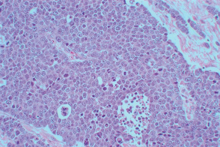
(A) CEA
(B) Chromogranin
(C) EMA
(D) Low molecular weight keratin
(E) p63
48. An endometrial biopsy from a 24-year-old woman is shown in Figure 15-21. The changes are most consistent with which secretory day?
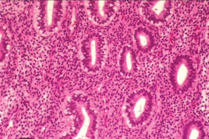
(A) Day 16
(B) Day 17
(C) Day 19
(D) Day 20
(E) Day 22
49. Predecidual change in the endometrium, limited to surrounding spiral arterioles, is characteristic of which secretory day?
(A) Day 21
(B) Day 22
(C) Day 23
(D) Day 24
(E) Day 25
50. Which of the following is the least likely cause of abnormal uterine bleeding in a 29-year-old woman?
(A) Anovulation
(B) Chronic endometritis
(C) Endometrial atrophy
(D) Endometrial polyp
(E) Leiomyoma
51. Which of the following features is considered characteristic of chronic endometritis?
(A) Eosinophils
(B) Granulomas
(C) Lymphocytes
(D) Lymphoid aggregates
(E) Plasma cells
52. The treatment of choice for chronic endometritis is which of the following?
(A) Antibiotics
(B) Curettage
(C) Hysterectomy
(D) No treatment required
(E) Oral contraceptives
53. All of the following are features of anovulation on an endometrial biopsy except
(A) Evidence of surface repair
(B) Fibrin thrombi in spiral arterioles
(C) Irregularly distributed and cystically dilated glands
(D) Patchy stromal breakdown
(E) Prominent thickened blood vessels
54. The risk of developing endometrial adenocarcinoma in the setting of isolated squamous morules is which of the following?
(A) Never
(B) <5%
(C) 5–10%
(D) 10–15%
(E) 20–25%
55. Eosinophilic metaplasia is most likely encountered in which of the following clinical settings?
(A) Adolescent with anovulatory cycles
(B) Menopausal woman on hormone replacement therapy
(C) Pregnancy
(D) Premenopausal woman with inadequate luteal phase
(E) Prepubertal girl with precocious puberty
56. All of the following are pregnancy-related changes of the uterus except
(A) Arias–Stella reaction
(B) Decidual cells
(C) Intermediate trophoblasts
(D) Nitabuch fibrin
(E) Papillary syncytial metaplasia
57. A 49-year-old woman has an endometrial biopsy that shows complex atypical hyperplasia. Her approximate risk of developing carcinoma is which of the following?
(A) <5%
(B) 5–10%
(C) 11–15%
(D) 16–20%
(E) 21–25%
58. Which of the following is not a risk factor for the development of endometrial hyperplasia?
(A) Anovulatory cycles
(B) Excess progesterone administration
(C) Obesity
(D) Polycystic ovarian syndrome
(E) Unopposed estrogen
59. Which of the following is the most common malignant tumor of the female genital tract?
(A) Cervical squamous cell carcinoma
(B) Endocervical adenocarcinoma
(C) Endometrial adenocarcinoma
(D) Ovarian serous carcinoma
(E) Uterine leiomyosarcoma
60. The pattern seen (Figure 15-22) in this endometrial tumor represents which variant of endometrial carcinoma?
(A) Clear cell
(B) Endometrial
(C) Mucinous
(D) Serous
(E) Transitional cell
61. Which of the following types of endometrial carcinoma has the worst prognosis?
(A) Clear cell
(B) Endometrioid
(C) Mucinous
(D) Squamous cell
(E) Transitional
62. All of the following are features of typical type I endometrial carcinoma except
(A) Hyperplasia precursor
(B) Mucinous carcinoma
(C) p53 alterations
(D) Premenopausal
(E) PTEN mutation
63. Which of the following parameters is used in grading an endometrial adenocarcinoma of the uterus?
(A) Amount of solid growth
(B) Depth of invasion
(C) Extent of squamous differentiation
(D) Number of mitotic figures
(E) Percent necrosis
64. All of the following features are useful in establishing the presence of squamous differentiation in an endometrial carcinoma except
(A) Discohesive cells
(B) Glassy cytoplasm
(C) Intercellular bridges
(D) Keratinization
(E) Sharp cell margins
65. A 46-year-old woman is diagnosed with a villo-glandular variant of endometrioid carcinoma of the uterus. All of the following are true regarding this variant except
(A) High-grade tumors
(B) May be associated with lymphovascular invasion
(C) Often shows areas of conventional endometrioid adenocarcinoma
(D) Second most common endometrioid carcinoma variant
(E) Villous architecture more common in superficial part of tumor
66. All of these features are characteristic of endometrioid adenocarcinoma of the uterus arising in the setting of hereditary nonpolyposis colon cancer syndrome (Lynch syndrome) except
(A) Lymphatic permeation
(B) MLH-1 positive staining
(C) Poor differentiation
(D) Prominent tumor infiltration by lymphocytes
(E) Sertoliform pattern
67. Psammoma bodies are most commonly seen in association with which of the following uterine tumors?
(A) Clear cell adenocarcinoma
(B) Mucinous adenocarcinoma
(C) Serous adenocarcinoma
(D) Small cell carcinoma
(E) Transitional cell carcinoma
68. Which of the following immunostains or special stains would be least useful in trying to differentiate endocervical versus endometrial adenocarcinoma?
(A) CEA
(B) Estrogen receptor
(C) Mucicarmine
(D) p16
(E) Vimentin
69. All of the following are features of the Arias–Stella reaction that can help in differentiating it from clear cell adenocarcinoma except
(A) Absent mitoses
(B) Hobnail cells
(C) Normal endometrial gland architecture
(D) Partial gland involvement
(E) Pseudonuclear inclusions
70. The tumor seen in this 53-year-old woman was biopsied in the uterus. The tumor stains with antibodies to cytokeratin 7, estrogen receptor, and progesterone receptor. Besides endometrioid carcinoma, what other lesion should be considered in the differential diagnosis? (Figure 15-23)
(A) Endometrial stromal sarcoma
(B) Leiomyosarcoma
(C) Metastatic breast carcinoma
(D) Metastatic colon cancer
(E) Placental site trophoblastic tumor
71. A 62-year-old woman is diagnosed with endometrioid adenocarcinoma of the uterus on biopsy. She undergoes a hysterectomy. Examination of the specimen shows tumor confined to the uterus and invading about 75% of the way through the myometrial wall. What stage is the tumor?
(A) Stage Ia
(B) Stage Ib
(C) Stage Ic
(D) Stage IIa
(E) Stage IIb
72. The uterine tumor shown in Figure 15-24 was resected in a 40-year-old woman. The tumor was subserosal in location and measured 3.4 cm in greatest dimension. The tumor is best classified as which of the following?
(A) Apoplectic leiomyoma
(B) Epithelioid leiomyoma
(C) Leiomyosarcoma
(D) Osteosarcoma
(E) Symplastic leiomyoma
73. The uterine mass illustrated in Figure 15-25 is best classified as which of the following?
(A) Apoplectic leiomyoma
(B) Leiomyoma with diffuse perinodular hydropic change
(C) Leiomyosarcoma
(D) Lipoleiomyoma
(E) Myxoid leiomyoma
74. Uterine leiomyomas are least likely to stain with which of the following antibodies?
(A) Actin
(B) Estrogen receptor
(C) h-caldesmon
(D) Oxytocin
(E) S-100 protein
75. Which of the following morphologic features is least consistent with a diagnosis of intravenous leiomyomatosis?
(A) Endothelium covered protrusions of smooth muscle
(B) Intersecting fascicles of desmin positivity
(C) Minimal cytologic atypia
(D) Minimal p53 immunoreactivity
(E) 6 Mitotic figures/10 high-power fields
76. The 2.6 cm uterine mass seen in this 38-year-old woman had up to 13 mitotic figures/10 high-power fields. The best diagnosis for this lesion is which of the following? (Figure 15-26)
(A) Cellular leiomyoma
(B) Epithelioid leiomyoma
(C) Gonadotropin-releasing hormone agonist treated leiomyoma
(D) Leiomyosarcoma
(E) Mitotically active leiomyoma
77. The most common site for benign metastasizing leiomyoma is which of the following?
(A) Bone
(B) Liver
(C) Lung
(D) Mediastinum
(E) Retroperitoneum
78. Which of the following is not a histologic feature associated with gonadotropin-releasing hormone agonist treatment of leiomyoma?
(A) Increased mitotic activity
(B) Infarct-like necrosis
(C) Lymphocyte infiltrate
(D) Nuclear crowding
(E) Smaller and fewer blood vessels
79. The lesion shown in Figure 15-27 was a 12 cm, partially necrotic mass arising in the uterus of a 56-year-old woman. The most likely diagnosis is which of the following?
(A) Bizarre leiomyoma
(B) Cellular leiomyoma
(C) Leiomyosarcoma
(D) Mitotically active leiomyoma
(E) Rhabdomyoma
80. PEComa of the uterus may stain with all of the following antibodies except
(A) CD10
(B) HMB-45
(C) Inhibin
(D) Microphthalmia transcription factor
(E) Melan-A
81. Which of the following antibodies is most commonly positive in the uterine mass shown in Figure 15-28.
(A) CD10
(B) Desmin
(C) CD99
(D) Melan-A
(E) Progesterone receptor
82. A 62-year-old woman presents with an enlarged uterus and pelvic pain. On hysterectomy, she is noted to have a 7 cm mass composed of a mixture of benign appearing endometrial glands and malignant stroma resembling fibrosarcoma. The best diagnosis for this lesion is which of the following?
(A) Endometrial polyp with cellular stroma
(B) Low-grade endometrial stromal sarcoma with sex cord-like differentiation
(C) Malignant mixed Müllerian tumor
(D) Müllerian adenofibroma
(E) Müllerian adenosarcoma
83. All of the following are risk factors for the development of malignant mixed Müllerian tumor of the uterus except
(A) Alcohol use
(B) Nulliparity
(C) Obesity
(D) Previous radiation
(E) Tamoxifen therapy
84. Median survival for malignant mixed Müllerian tumors of the uterus is best approximated by which of the following?
(A) About 1 year
(B) About 2 years
(C) About 5 years
(D) About 8–10 years
(E) <6 months
85. Peutz–Jeghers syndrome is associated with the development of which of the following changes in the fallopian tube?
(A) Decidual change
(B) Mucinous metaplasia
(C) Pseudocarcinomatosis hyperplasia
(D) Salpingitis isthmica nodosa
(E) Walthard cysts
86. All of the following are risk factors for the development of a tubal ectopic pregnancy except
(A) Decidual metaplasia
(B) Endometriosis
(C) History of salpingitis
(D) Pelvic inflammatory disease
(E) Prior tubal surgery
87. Which of the following organisms is least commonly associated with pelvic inflammatory disease?
(A) Anaerobic bacteria
(B) Chlamydia
(C) Mycoplasma
(D) Neisseria
(E) Staphylococcus
88. Bilateral salpingitis marked by caseating mucosal granulomas, chronic inflammation, and fibrosis in the muscularis propria and a paucity of eosinophils is most characteristic of which of the following?
(A) Crohn disease
(B) Parasitic infection
(C) Reaction to lipoidal contrast agents
(D) Sarcoidosis
(E) Tuberculosis
89. A 1.2 cm subserosal mass is identified in the fallopian tube of a 51-year- old woman. The lesion, shown in Figure 15-29, is best classified as which of the following?
(A) Adenocarcinoma
(B) Adenomatoid tumor
(C) Female adnexal tumor of probable Wolffian duct origin
(D) Leiomyoma
(E) Lymphangioma
90. The tumor shown in Figure 15-30 is the same lesion as in Question 89. The positive staining shown most likely is associated with which of the following antibodies?
(A) Calretinin
(B) CD15
(C) CEA
(D) Factor VIII
(E) TAG-72
91. Which of the following fallopian tube lesions is most frequently associated with BRCA1or BRCA2 mutations?
(A) Adenocarcinoma
(B) Adenomatoid tumor
(C) Female adnexal tumor of probable Wolffian origin
(D) Metaplastic papillary tumor
(E) Papilloma
92. All of the following are true regarding a female adnexal tumor of probable Wolffian origin except
(A) Commonly arise in the broad ligament
(B) Low-grade tumor
(C) May have tubular or sieve-like growth patterns
(D) Stains with CEA
(E) Stains with cytokeratins AE1/AE3
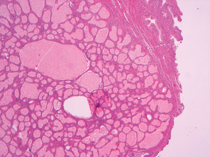
93. All of the following are features or findings commonly associated with the ovarian changes shown in Figure 15-31 except
(A) Anovulation
(B) Hirsutism
(C) Infertility
(D) Insulin resistance
(E) Postmenopausal peak
94. A 59-year-old woman had endometrial carcinoma diagnosed on a hysterectomy. On examination of the ovaries removed at the same time, the change shown in Figure 15-32 was noted. The best diagnosis for this lesion is which of the following?
(A) Decidual change
(B) No pathologic change
(C) Steroid cell tumor
(D) Stromal hyperthecosis
(E) Stromal luteoma
95. Multiple circumscribed, red-brown ovarian nodules presenting during pregnancy most likely represent which of the following?
(A) Leydig cell hyperplasia
(B) Leydig cell tumors
(C) Luteinized follicular cysts
(D) Pregnancy luteomas
(E) Stromal hyperplasia
96. A 19-year-old woman presents with abdominal pain and a 12 cm right ovary that is excised and shown in Figure 15-33. The lesion most likely represents which of the following?
(A) Hyperreactio luteinalis
(B) Lymphangioma
(C) Massive ovarian edema
(D) Myxoid leiomyoma
(E) Pregnancy luteoma
97. The most common surface epithelial stromal tumor type of the ovary is which of the following?
(A) Clear cell
(B) Endometrioid
(C) Mucinous
(D) Serous
(E) Transitional
98. The overall 5-year survival for serous borderline tumor of the ovary is best approximated by which of the following
(A) 40–50%
(B) 60%
(C) 70%
(D) 80%
(E) 90%
99. Which of the following immunostains would positively stain a well-differentiated papillary mesothelioma and allow its differentiation from serous borderline tumor?
(A) CA125
(B) Calretinin
(C) BerEP4
(D) CD15
(E) Cytokeratin 7
100. The ovarian tumor shown in Figure 15-34 arose in a 46-year-old woman and is best classified as which of the following?
(A) Borderline mucinous tumor
(B) Endometrioid carcinoma
(C) Mucinous adenocarcinoma
(D) Mucinous cystadenoma
(E) Metastatic appendicial carcinoma
101. Mucinous epithelial tumors of the ovary may coexist with all of the following tumors except
(A) Brenner tumor
(B) Carcinoid tumor
(C) Granulosa cell tumor
(D) Mature cystic teratoma
(E) Sertoli–Leydig tumor
102. Primary mucinous adenocarcinomas of the ovary demonstrate positive immunostaining with all of the following antibodies except
(A) CA125
(B) CDX2
(C) CEA
(D) Cytokeratin 7
(E) Cytokeratin 20
103. All of the following are true regarding endometrioid carcinomas arising in the ovary except
(A) About 15–20% of patients have a synchronous primary endometrial adenocarcinoma.
(B) They demonstrate ER and PR immuno-reactivity.
(C) Most patients present in the 5th-7th decades.
(D) Most present as bilateral ovarian masses.
(E) Serum CA125 levels are commonly elevated.
104. All of the following are true regarding clear cell carcinoma of the ovary except
(A) It is associated with paraneoplastic hyper-calcemia.
(B) It is associated with pelvic endometriosis.
(C) Most occur in nulliparous women.
(D) Patients with this tumor are at risk of developing pelvic venous thromboses.
(E) Stage for stage, clear cell carcinoma has a better prognosis than serous carcinoma.
105. The right ovarian tumor illustrated in Figure 15-35 presented in a 49-year-old woman. The best diagnosis is which of the following?
(A) Benign Brenner tumor
(B) Borderline Brenner tumor
(C) Granulosa cell tumor
(D) Malignant Brenner tumor
(E) Transitional cell carcinoma
106. Which of the following ovarian tumors is associated with Gorlin syndrome?
(A) Adult granulosa cell tumor
(B) Fibroma
(C) Sertoli–Leydig cell tumor
(D) Sex cord tumor with annular tubules
(E) Thecoma
107. A 52-year-old woman presents with a left ovarian mass and a recent history of endometrial hyper-plasia. The lesion illustrated in Figure 15-36 stains with antibodies to inhibin and CD10 and shows cytoplasmic oil-red-O staining. The best diagnosis for this tumor is which of the following?
Stay updated, free articles. Join our Telegram channel

Full access? Get Clinical Tree


