, Tsunehisa Kaku2, Toru Sugiyama3 and Steven G. Silverberg4
(1)
Matsue City Hospital, Matsue, Shimane, Japan
(2)
Department of Health Sciences, Department of Health Sciences Graduate School of Medical Sciences, Fukuoka, Fukuoka, Japan
(3)
Department of Obstetrics and Gynecology, Iwate Medical University School of Medicine, Morioka, Iwate, Japan
(4)
Department of Pathology, University of Maryland School of Medicine, Baltimore, MD, USA
2.1 Gross Pathology
There is no distinctive gross appearance of ovarian clear cell carcinoma (CCC), which usually is recognizable as malignant but not specifically as CCC. Most of these tumors are solid, soft to firm, and often multinodular, often (especially if large) with foci of necrosis and/or hemorrhage. The color may vary from yellow to white to brown (Fig. 2.1).
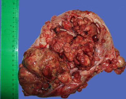
Fig. 2.1
Clear cell carcinoma gross appearance. Large soft multinodular red-gray tumor invading through its capsule. The appearance is malignant but otherwise nonspecific
A gross finding in some tumors which suggests CCC is one or more foci of tumor within or adjacent to a hemorrhagic smooth-walled cyst with the appearance of endometriosis (Figs. 2.2, 2.3, and 2.4). Most malignant ovarian tumors with this appearance are either CCC or endometrioid adenocarcinoma, with a slight preponderance of the former.
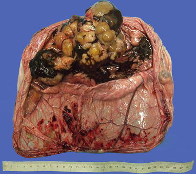
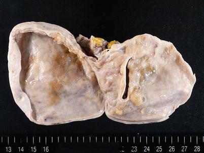
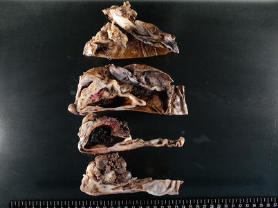
Fig. 2.2
Hemorrhagic smooth-walled endometriotic cyst containing masses of brown necrotic tumor
Fig. 2.3
Small solid gray-white tumor foci (R) in cyst lined focally by golden-brown altered blood, representing origin in endometriosis (formalin-fixed specimen)
Fig. 2.4
Formalin-fixed and sectioned blood-filled endometriotic cyst surrounded by gray-brown multinodular tumor which is also present in the cyst lumen
A somewhat different macroscopic appearance is present in those ovarian CCCs of the adenofibromatous type (cf. Chap. 3). Among our cases, 8–9 % expressed this phenotype, of which approximately half were predominantly adenofibromatous. These tumors are usually not obviously grossly malignant and may resemble Brenner tumors or adenofibromas, with a predominantly firm solid architecture punctuated by small or large cystic spaces.
Stay updated, free articles. Join our Telegram channel

Full access? Get Clinical Tree


