glycosides, glucosinolate compounds, cysteine derivatives and miscellaneous glycosides

Chapter 25 Cyanogenetic glycosides, glucosinolate compounds, cysteine derivatives and miscellaneous glycosides


In addition to the important groups of glycosides discussed in previous chapters, there are a number of other groups of some medicinal interest. Two of these, the cyanogenetic glycosides and the glucosinolate compounds, are characteristic of certain groups of plants and have similarities in their biosynthetic origins.


CYANOGENETIC GLYCOSIDES


The poisonous properties of the roots of Manihot utilissima (cassava) have long been known to primitive tribes; they use it as an important foodstuff, having first found methods to remove its poison. In 1830 the cyanogenetic glycoside manihotoxin was isolated from it, and in the same year amygdalin was obtained from bitter almonds, linamarin from linseed and phaseolunatin from a bean, Phaseolus lunatus. These yield prussic acid on hydrolysis and were the first discovered cyanogenetic or cyanophoric glycosides. Over 2000 plant species involving about 110 families are estimated to be cyanogenetic. Professor Lindley, a teacher of pharmaceutical students in London, realized as early as 1830 that the presence or absence of HCN was of taxonomic importance and used it as a character for separating the subfamilies of the Rosaceae. At the species level the presence or absence of prussic acid may denote varieties or different chemical races of the same species (e.g. Prunus amygdalus yields both bitter and sweet almonds). Interest in cyanogenetic principles as chemotaxonomic characters continues to receive much attention, as does the general biochemistry of cyanide in plants and microorganisms.


Many of these glucosides, but not all, are derived from the nitrile of mandelic acid. Although they contain nitrogen their structure is that of O-and not N-glycosides. The sugar portion of the molecule may be a monosaccharide or a disaccharide such as gentiobiose or vicianose. If a disaccharide, enzymes present in the plant may bring about hydrolysis in two stages, as in the case of amygdalin (amygdaloside), Fig. 25.1.


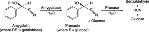
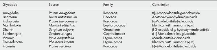
Table 25.1 gives some well-known cyanogenetic glycosides isolated from various sources between 1830 and 1907.


Tests


To test for a cyanogenetic glycoside qualitatively the material is well broken and placed in a small flask with sufficient water to moisten. In the neck of the flask a suitably impregnated strip of filter-paper is suspended by means of a cork. The paper may be treated in either of the following ways to give a colour reaction with free hydrocyanic acid. Either sodium picrate (yellow), which is converted to sodium isopurpurate (brick-red), or a freshly prepared solution of guaiacum resin in absolute alcohol which is allowed to dry on the paper and treated with very dilute copper sulphate solution. The latter test-paper turns blue with prussic acid. If the enzymes usually present in the material have not been destroyed or inactivated, the hydrolysis takes place within about an hour when the flask is kept in a warm place. More rapid hydrolysis will result if a little dilute sulphuric acid is added and the flask gently heated. The depth of colour produced with sodium picrate paper can be used for semiquantitative evaluations.


For materials containing a fairly high percentage of cyanogenetic glycosides (e.g. bitter almonds) the amount may be determined quantitatively by placing the plant in a flask with water and tartaric acid and passing steam through until all the hydrocyanic acid has distilled into a receiver. The distillate is then adjusted to a definite volume and aliquots titrated with standard silver nitrate solution. More sensitive methods including the direct determination of individual glycosides by GLC of their TMS derivatives are now available.


Biogenesis


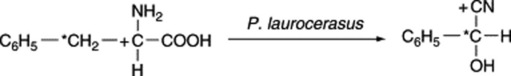
The aglycones of cyanogenetic glycosides are derived solely from nitrogen intermediates. The biosynthesis of prulaurasin (DL-mandelonitrile glucoside) has been studied in the leaves of Prunus laurocerasus. Phenyl[3-14C]alanine, phenyl[2-14C]alanine and phenyl[1-14C]alanine were fed to the leaves and the hydrolytic products of the isolated glycosides were examined. The three labelled precursors gave, respectively, active benzaldehyde and inactive hydrocyanic acid; inactive benzaldehyde and active hydrocyanic acid; and inactive benzaldehyde and active hydrocyanic acid; and inactive hydrolytic products consistent with the following incorporation:


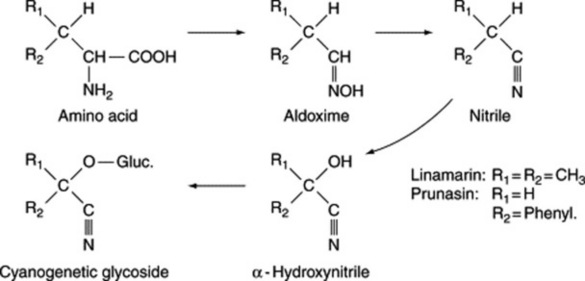
Similarly, phenyl[2-14C]alanine fed to P. amygdalus gives amygdalin with most activity in the carbon atom of the nitrile. Experiments with doubly labelled amino acids have shown that the nitrile nitrogen of the cyanogen is derived from the nitrogen atom of the amino acid. Similar results have been obtained with dhurrin isolated from sorghum seedlings fed with labelled tyrosine. More recent work has sought to determine the nature of the intermediates involved in the above conversions and, for prunasin and linamarin, the participation of oximes and nitriles has been demonstrated (Fig. 25.2).


For a report of a lecture on the biosynthesis, compartmentation and catabolism of cyanogenetic glycosides including amygdalin, linamarin and lotaustralin see E. E. Conn, Planta Med., 1991, 57 (Suppl. Issue No 1), SI. Nahrstedt (Proc. Phytochem. Soc. Europe, 1992, 33, 249) reviewed (84 refs) progress concerning the biology of cyanogenetic glycosides.


A review (107 refs) asking ‘Why are so many plants cyanogenetic?’ (D. A. Jones, Phytochemistry, 1998, 47, 155) illustrates the continuing interest in these plants, an interest which is, however, largely non-pharmaceutical.


Wild cherry bark


Wild cherry bark (Wild Black Cherry or Virginia Prune Bark; Prunus Serotina) is the dried bark of Prunus serotina (Rosaceae). The plant is a shrub or tree widely distributed in Canada and the USA, extending from Ontario to Florida and westward to Dakota and Texas. Commercial supplies are obtained from Virginia, North Carolina and Tennessee. The most esteemed bark is collected in the autumn, at which time it is most active. After careful drying it should be kept in airtight containers.


Macroscopical characters


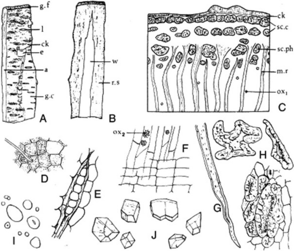
The drug usually occurs in curved or channelled pieces up to 10 cm long, 5 cm wide and 0.3–4.0 mm thick (Fig. 25.3). Much larger pieces of trunk bark, up to 8 mm thick, may be found but the BP (1980) maximum thickness is 4.0 mm and is known commercially as ‘Thin Natural Wild Cherry Bark’. The branch bark, if unrossed, is covered with a thin, glossy, easily exfoliating, reddish-brown to brownish-black cork, which bears very conspicuous whitish lenticels. In the rossed bark pale buff-coloured lenticel scars are seen and the outer surface is somewhat rough, some of the cortex having been removed and the phloem exposed. The inner surface is reddish-brown and has a striated and reticulately furrowed appearance, which is caused by the distribution of the phloem and medullary rays. Patches of wood sometimes adhere to the inner surface. The drug breaks with a short, granular fracture. When slightly moist it has an odour of benzaldehyde. Taste is astringent and bitter.


< div class='tao-gold-member'>

Stay updated, free articles. Join our Telegram channel

Jul 18, 2016 | Posted by in PHARMACY | Comments Off on glycosides, glucosinolate compounds, cysteine derivatives and miscellaneous glycosides

Full access? Get Clinical Tree

Get Clinical Tree app for offline access