Glomus Tumor
Key Facts
Terminology
Synonyms: Glomangioma, glomic tumor
Clinical Issues
Cough
Shortness of breath
Asymptomatic
Image Findings
Coin lesion in intrapulmonary location
Central tumor obstructing bronchial lumen
Microscopic Pathology
Solid and homogeneous cellular proliferation
Ectatic blood vessels
Cellular proliferation with clear cytoplasm mimicking “fried-egg” appearance
Mitotic figures are absent
Necrosis and hemorrhage are absent
Top Differential Diagnoses
Glomangiosarcoma
Mitotic figures and cellular pleomorphism are most important features to separate from glomangioma
Leiomyoma
Rarely displays prominent ectatic blood vessels with edema of wall
Both tumors may show similar immunohistochemical profile
Tumor cells are mostly oval or spindled
Carcinoma
Displays more cellular atypia and mitotic activity
Shows positive staining for epithelial markers
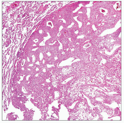 Low-power view of a primary pulmonary glomus tumor shows a well-defined tumor mass replacing normal lung parenchyma. |
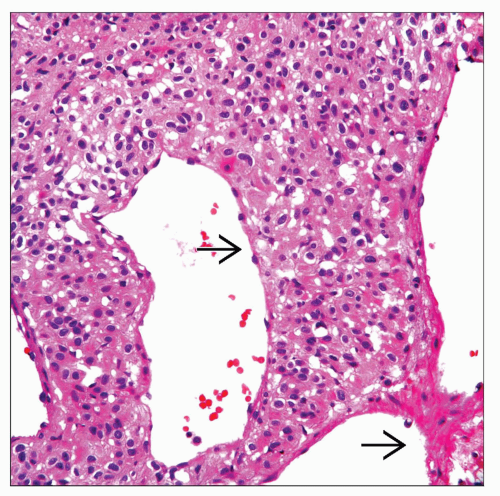 High-power view of a primary pulmonary glomus tumor shows ectatic blood vessels  and a cellular proliferation composed of medium-sized cells with clear and eosinophilic cytoplasm. and a cellular proliferation composed of medium-sized cells with clear and eosinophilic cytoplasm. |
TERMINOLOGY
Synonyms
Glomangioma, glomic tumor
Definitions
Benign tumor with smooth muscle differentiation
ETIOLOGY/PATHOGENESIS
Etiology
Glomus tumors are believed to originate from glomus body
Debated whether it represents a true tumor or hyperplasia
CLINICAL ISSUES
Epidemiology
Incidence
Very rare tumor in lung
Age
Cases reported have been in adults
Gender
No gender predilection
Presentation
Cough
Shortness of breath
Asymptomatic
Treatment
Surgical approaches
Complete surgical resection
Prognosis
Excellent
IMAGE FINDINGS
General Features
Coin lesion in intrapulmonary location
Central tumor obstructing bronchial lumen
MACROSCOPIC FEATURES
General Features
Well-circumscribed tumor embedded in lung parenchyma
White to tan in color without hemorrhage &/or necrosis
Size
May vary from 1-5 cm in diameter
MICROSCOPIC PATHOLOGY
Histologic Features
Well-circumscribed tumor nodule
Solid and homogeneous cellular proliferation
Ectatic blood vessels
Cellular proliferation with clear cytoplasm mimicking “fried-egg” appearance
Hemangiopericytic pattern
Mucohyaline changes
Mitotic figures are absent
Necrosis and hemorrhage are absent
Predominant Pattern/Injury Type
Solid
Predominant Cell/Compartment Type
Smooth muscle
DIFFERENTIAL DIAGNOSIS


