55 Glaucoma
Pathophysiology
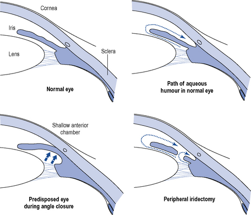
The physiological precipitating factors of PACG in predisposed eyes are not fully understood. Two theories currently exist. The dilator muscle theory suggests that contraction of the dilator muscle causes a posterior movement, which increases the apposition between the iris and anteriorly located lens and the degree of physiological pupillary block. The simultaneous dilation of the pupil renders the peripheral iris more flaccid, and causes the pressure in the posterior chamber to increase and the iris to bow anteriorly. Eventually, the peripheral iris obstructs the angle and the IOP rises (Fig. 55.1).
Investigations
IOP may be measured by tonometry, such as indentation tonometry in which a plunger is applied to the cornea and the amount of indentation on the eye reflects the pressure within it. Tonography is a technique used to measure the outflow of aqueous humour from the eye, resulting from indentation of the eye, using a tonometer. Gonioscopy is used to estimate the width of the chamber angle, with the aid of a slit lamp. Perimetry is important for both the diagnosis and management of glaucoma by detecting early scotomata and larger changes in visual field. Other investigations which should be routinely offered to patients include a central corneal thickness measurement (CCT), as this has an effect on the measured IOP and may affect the efficacy of certain drug treatments (Johnson et al., 2008), and fundus examination for optic nerve assessment with dilatation using stereoscopic slit-lamp biomicroscopy. There are also guidelines for the monitoring of these patients and at what intervals their IOP, optic nerve head and visual fields should be checked (National Institute for Health and Clinical Excellence, 2009).
Treatment
Chronic open-angle glaucomas
The initial treatment of COAG is usually medical. Topical administration is the preferred type of therapy, and there is a wide range of preparations available (Table 55.1). The chosen drug should be administered at its lowest concentration and as infrequently as possible to obtain the desired effect over the whole 24-h period as a high level of diurnal variation in IOP has been shown to be a more important factor in visual field development than the average IOP. A drug with few potential side effects should be chosen, with oral therapy retained as the final option. The prostaglandin analogues latanoprost and travoprost and the prostamide bimatoprost are indicated for first-line use, as are the β-blockers as these produce the greatest fall in IOP (Table 55.2). Carbonic anhydrase inhibitors and sympathomimetics, which result in a smaller fall in IOP, are reserved for patients unresponsive to first-line drugs, in patients in whom the first-line agents are contraindicated or as adjunctive therapy.
Table 55.1 Drugs used in the treatment of chronic open-angle glaucoma
| Therapeutic category | Primary mechanism |
|---|---|
| Topical prostaglandins | Increase aqueous outflow |
| Topical prostamides | Increase aqueous outflow |
| Topical β-blocking agents | Decrease aqueous formation |
| Topical miotics | Increase aqueous outflow |
| Topical adrenergic agonists | Increase aqueous outflow and decrease aqueous formation |
| Topical carbonic anhydrase inhibitors | Decrease aqueous formation |
| Oral carbonic anhydrase inhibitors | Decrease aqueous formation |
Table 55.2 Comparison of reduction in intraocular pressure (IOP) with a range of ocular hypotensive drugs (adapted from van der Valk et al., 2009)
| Drug | Relative IOP change from baseline at trough (%) (95% CI) | Relative IOP change from baseline at peak (%) (95% CI) |
|---|---|---|
| Bimatoprost | −28 (−29 to −27) | −33 (−35 to −31) |
| Travoprost | −29 (−32 to −25) | −31 (−32 to −29) |
| Latanoprost | −28 (−30 to −26) | −31 (−33 to −29) |
| Timolol | −26 (−28 to −25) | −27 (−29 to −25) |
| Betaxolol | −20 (−23 to −17) | −23 (−25 to −22) |
| Dorzolamide | −17 (−19 to −15) | −22 (−24 to −20) |
| Brinzolamide | −17 (−19 to −15) | −17 (−19 to −15) |
| Brimonidine | −18 (−21 to −14) | −25 (−28 to −22) |
| Placebo | −5 (−9 to −1) | −5 (−10 to 0) |
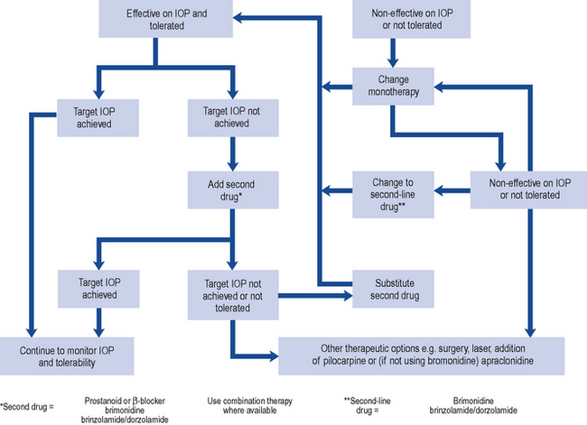
In most cases, the initial topical treatment is with a prostaglandin analogue or prostamide. If this is ineffective, another prostaglandin or prostamide may be substituted or a β-blocker used instead. Patients not reaching their target pressure on one first-line agent may be prescribed another concomitantly. Alternatively, a carbonic anhydrase inhibitor or a sympathomimetic may be added to one of the first-line drugs. Guidance on the treatment of people with OHT or suspected COAG has been published (National Institute for Health and Clinical Excellence, 2009). The treatment options (Table 55.3) take into account central corneal thickness and age, although age thresholds are only appropriate where vision is normal (OHT with or without suspected COAG) and the treatment is purely preventative. Pilocarpine is no longer recommended by NICE but is recommended as a second-line agent in European guidelines (European Glaucoma Society, 2008). Oral therapy with carbonic anhydrase inhibitors is usually reserved for use as the final stage of treatment in those complex glaucomas awaiting surgery (Fig. 55.2).
Table 55.3 Treatment of people with ocular hypertension or suspected COAG (National Institute for Health and Clinical Excellence, 2009)


Fig. 55.2 Topical therapy for chronic open-angle glaucoma (COAG) based on recommendations of European Glaucoma Society (2008).
It has been proposed that the improved control of IOP with newer therapies has led to a reduction in surgery for glaucoma (Kenigsberg, 2007). However, some patients still require surgical intervention to reach their target pressure.
The most frequently performed surgical procedures create a fistula to act as a new route for aqueous outflow and the most frequently performed surgery of this type, trabeculectomy, appears more effective in controlling IOP than laser trabeculoplasty, in which a series of laser burns is applied to the trabecular meshwork to improve the outflow of aqueous humour. (Rolim de Moura et al., 2007).
Primary angle-closure glaucoma
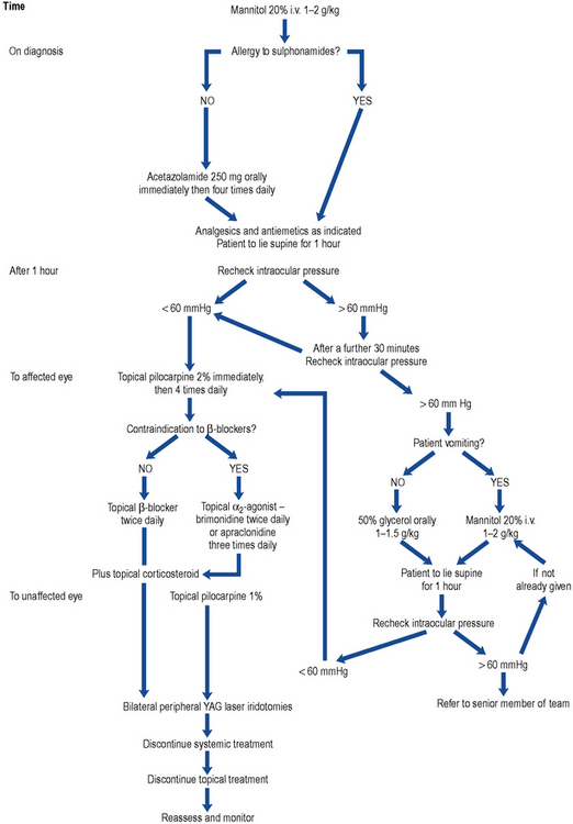
The medical management of acute PACG is essentially to prepare the eye for surgical treatment. The aim of treatment is to decrease the IOP and associated inflammation. Analgesics and antiemetics are sometimes needed, dependent on symptom severity, to make the patient comfortable. It is usual to treat the unaffected eye prophylactically with miotics (Fig. 55.3).

Fig. 55.3 Algorithm for the treatment of primary angle-closure glaucoma (PACG). YAG, yttrium aluminium garnet.
Once the IOP has been reduced medically, the condition is treated surgically, by either surgical peripheral iridectomy or laser iridotomy, to remove an area of the peripheral iris to allow flow of aqueous humour through an alternative pathway (see Fig. 55.1). Filtration surgery is indicated if a large proportion of the angle has been permanently closed by adhesions between the iris and the cornea.
Drug treatment
Ocular prostanoids: prostaglandin analogues and prostamides
The National Institute for Health and Clinical Excellence (2009) guideline does not differentiate between the prostaglandin analogues and the prostamide bimatoprost, but recommends one of this class for the treatment of COAG, certain patients with OHT and certain COAG suspects (see Table 55.4)
The prostaglandin analogues, latanoprost, travoprost and tafluprost, are ester compounds thought to achieve a fall in IOP primarily by increasing the uveoscleral outflow with no significant effect on other parameters of aqueous humour dynamics, while the prostamide bimatoprost is thought to increase outflow through both trabecular and uveoscleral outflow pathways. However, there is still debate about the precise mechanisms of action of this group (Lim et al., 2008; Toris et al., 2008).
In a meta-analysis, these drugs have been shown to have a greater effect on both peak and trough readings of IOP than timolol, betaxolol, dorzolamide, brinzolamide or brimonidine, producing falls in IOP of 28–29% at trough and 31–33% at peak as shown in Table 55.2 (van der Valk et al., 2009).
Randomised head-to-head evaluations of prostaglandin therapy demonstrate similar efficacy, but differing hyperemia effects with bimatoprost and travoprost causing more hyperaemia than latanoprost (Eyawo et al., 2009; Honrubia et al., 2009). However, these studies were conducted before the introduction of the new formulations of bimatoprost 0.01% and the benzalkonium chloride–free travoprost.
The prostanoids have some interesting local side effects (Table 55.5). Pigmentation of the iris occurs in patients with mixed colour (green-brown or blue-brown) irides after 3–6 months of use and is a result of increased deposition of melanin in the melanocytes. An increase in the length and thickness of the eyelashes and pigmentation of the palpebral skin are also known side effects. Use of these drugs may lead to disruption of the blood–aqueous barrier in patients with aphakia and pseudophakia (no or false lens, respectively) and increase the risk of developing cystoid macular oedema; they should be used with caution in such patients. Reactivation of herpes simplex infection has been reported with bimatoprost, travoprost and latanoprost.
Table 55.5 Side effects of prostanoids
| Ocular | Systemic |
|---|---|
| Asthenopia | Abdominal cramp |
| Allergic conjunctivitis | Asthenia |
| Blepharitis | Asthma |
| Blepharospasm | Bradycardia |
| Browache | Dizziness |
| Cataract | Dyspnoea |
| Conjunctival follicles, papillae | Elevated liver function |
| Conjunctival hyperaemia | Headache |
| Cystoid macular oedema | Hirsutism |
| Corneal erosion | Hypertension |
| Conjunctival oedema | Hypotension |
| Deepening of lid sulcus | Infection (primarily URTIs) |
| Distichiasis | Peripheral oedema |
| Eye discharge | Skin rash |
| Eye pain | |
| Eyelash changes – increased number, length, thickness, pigmentation, misdirection, poliosis | |
| Eyelid oedema, eyelid retraction | |
| Eyelid and periocular skin darkening | |
| Foreign body sensation | |
| Increase in vellus hair on eyelids | |
| Increased iris pigmentation | |
| Iritis | |
| Lid margin crusting | |
| Localised skin reactions on the eyelids | |
| Ocular burning, dryness, irritation pruritus, fatigue | |
| Photophobia | |
| Punctate epithelial erosions | |
| Retinal haemorrhage | |
| Tearing | |
| Uveitis | |
| Visual disturbance |
URTIs, upper respiratory tract infection
Latanoprost
Latanoprost, which is converted to its active free acid on entering the eye, was the first of the prostanoids to be launched and is the market leader. Like timolol amongst the β-blockers, latanoprost is the drug in this class against which new drugs or combinations are assessed. It must be administered in the evening for maximum effect (Stewart et al., 2008). Latanoprost is generally very well tolerated. Serious adverse drug reactions were reported in only 17/3936 (0.43%) patients using latanoprost over a 5-year period (Goldberg et al., 2008), and patient persistence with latanoprost therapy is better than that with all other frequently used monotherapies (Rahman et al., 2009). Latanoprost is not heat stable and requires refrigeration; however, it is stable enough to be stored at room temperature for the 4-week in-use period applied in the UK. The concentration of the preservative benzalkonium chloride in latanoprost eye drops and bimatoprost 0.01% may prevent their use in certain patients. The benzalkonium chloride concentration in these formulations is 0.02%, which is four times that in bimatoprost 0.03% eye drops. The current formulation of travoprost, launched in January 2011, contains polyquaternium-1 rather than benzalkonium chloride.
Travoprost
Patients treated with travoprost show good diurnal fluctuation control, and an ocular hypotensive effect was shown to last for over 3 days. Patients previously treated with β-blockers, α2-agonists, carbonic anhydrase inhibitors, latanoprost and the dorzolamide-timolol fixed combination respond to travoprost. Administration time is not as important with travoprost as with latanoprost as no statistical difference was noted in the percentage fall in IOP whether travoprost was administered in the morning or in the evening (Stewart et al., 2008). Travoprost is a stable compound throughout a range of temperatures, and the commercially available product does not require refrigeration.
Tafluprost
Tafluprost is the first of the prostanoids available in a preservative-free form. In terms of efficacy and safety, it has been shown to be equivalent to a preserved preparation of tafluprost (Hamacher et al., 2008) and useful in patients allergic to, or intolerant of, benzalkonium chloride.
Tafluprost as adjunctive therapy to timolol results in consistently greater reductions in IOP compared with vehicle in patients with glaucoma or OHT that is inadequately controlled with timolol monotherapy (Egorov and Ropo, 2009).


