Chapter 12 Genitourinary System
Kidney – Structure and Function
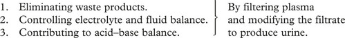
The kidney has several functions:
Many substances will show altered values in renal failure, but the following are always elevated and are commonly used in estimating the progress of the disease.

In practice renal function is now commonly assessed by estimated glomerular filtration rate (eGFR), which combines serum creatinine with the age and sex of patient.
Chronic Kidney Disease can be classified as follows:
CKD | Stage | eGFR/mL/min/1.73m2 |
---|---|---|
1 | with an abnormality* | >90 |
2 | with an abnormality* | 60–89 |
3 | 30–59 | |
4 | 15–29 | |
5 | <15 |
The structure and function and diseases will be discussed separately, but diseases of one may well affect others.
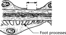
Glomerular Structure and Function
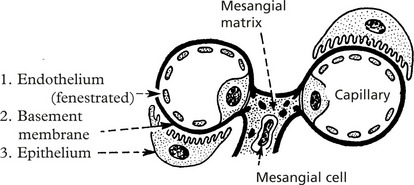
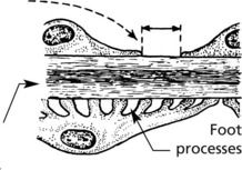
The glomerulus, of which there are over 600000 in the adult, consists of an invagination of a capillary network, derived from the afferent arteriole, into Bowman’s capsule – the beginning of the proximal tubule.
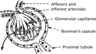
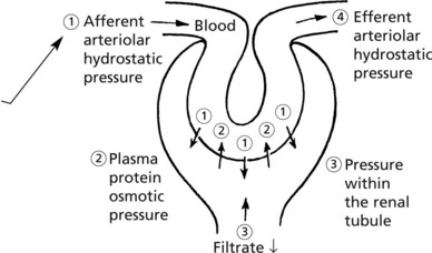
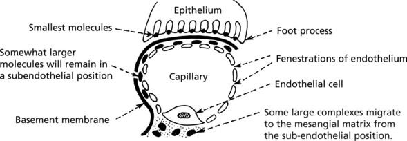
The glomerulus is an efficient filter due to the large surface area of the glomerular capillaries.
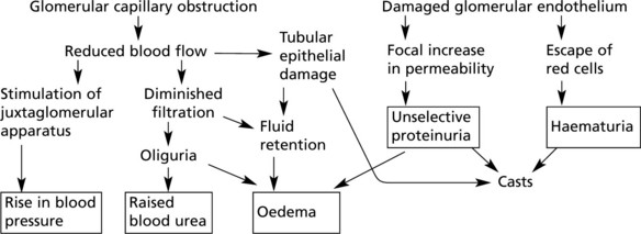
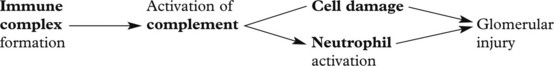
Ultrastructure
The barrier separating blood from the lumen of the matrix nephron consists of 3 layers:
Mesangial cells have three functions:
These layers form a complicated sieve controlling glomerular permeability. Wide pores (70–100 nm) in the endothelium allow all components to reach the basement membrane.
The basement membrane produced by the endothelium and epithelium has a strong anionic (negative) charge which repels major plasma proteins (also anionic). It has a central dense layer (lamina densa).
The epithelial cells are attached to the basement membrane by foot processes, separated by ‘slit pores’ 30–60 nm in diameter.
Filtration
The glomerular filtrate is virtually protein free plasma. Its rate of production is dependent upon:
Variation in any of these factors affects the output of urine. Diminished urinary output results from a reduction in renal blood flow as in shock, from an increase in osmotic pressure as in haemoconcentration, or from obstruction to the outflow of urine.
Glomerular Diseases
The mechanisms underlying these changes are as follows:
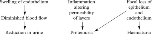
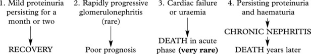
These lead to 4 main clinical syndromes:
The main forms of glomerular disease are described in the following pages.
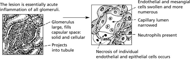
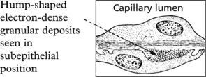
Acute Diffuse Proliferative Glomerulonephritis
This disease classically follows 2–3 weeks after an infection – usually pharyngitis due to Group A haemolytic streptococci. It is commonest in children and young adults who develop the NEPHRITIC SYNDROME: oliguria, proteinuria, haematuria (urine is smoky and dark), moderate hypertension and facial (periorbital) oedema. This disease is now uncommon in fully developed countries.
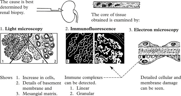
Immune complexes are identified by:
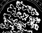
The clinical findings can be correlated with pathology as follows:
Prognosis – The disease usually resolves in 1–2 weeks, particularly in children, but in adults complications are more common.
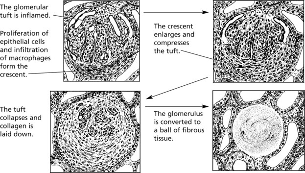
Crescentic (Rapidly Progressive) Glomerulonephritis
Without treatment, this disease progresses to end-stage renal failure in weeks or months. The histological hallmark is crescent formation in >50% of glomeruli.
Many types of glomerulonephritis can progress to crescentic GN. Examples include:
In this serious disorder there is both renal and often pulmonary damage. Immunofluorescence shows the cause – an antibody to type IV collagen in the glomerular basement membrane (anti-GBM) which also damages pulmonary alveolar membranes.
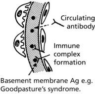
IF shows linear deposits of IgG and C3 in the capillary basement membranes
Prognosis – Without treatment, most patients die within 6 months.
Immunosuppressive drugs (e.g. steroids, cyclophosphamide) and plasma exchange to (remove anti-GBM antibodies) improve the prognosis but many patients require dialysis or transplantation. Hypertension is a further serious complication.
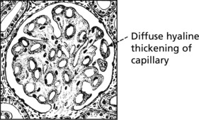
Membranous Glomerulonephritis
This accounts for around 30% of cases of the nephrotic syndrome in adults; some patients present with asymptomatic proteinuria.
Aetiology
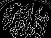
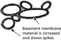
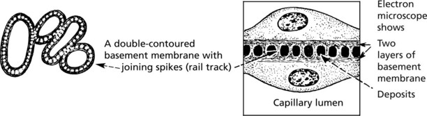
Pathology – There is generalised thickening of capillary basement membrane.
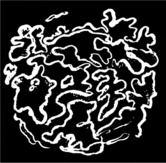
Specific silver staining of the basement membrane shows a typical pattern.
Immunofluorescence reveals deposition of IgG in the capillary walls.
Electron microscope findings explain this appearance.
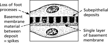
In the later stages there is a massive increase in basement membrane material.
Tubular changes – In the early stages, protein droplets and lipid globules appear in the tubular epithelium. If the disease progresses there is atrophy of the tubules and interstitial fibrosis.
Prognosis – In about 25% of patients the disease remits spontaneously, the remainder continuing to have proteinuria and in 40% chronic renal failure eventually supervenes.
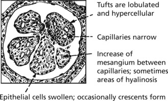
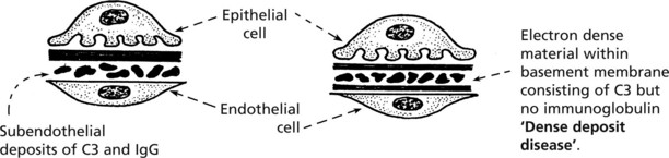
Mesangiocapillary (Membranoproliferative) Glomerulonephritis
In this form of glomerulonephritis there is an increase both in cells and mesangial matrix within glomeruli.
Silver staining of basement membrane
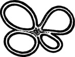
Duplication of basement membrane as in membranous glomerulonephritis, but without spikes.
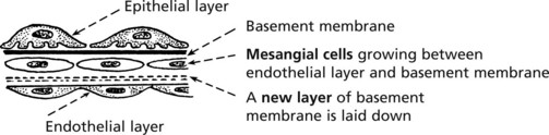
These changes are due to ‘mesangial interposition’
Using electron microscopy and immunofluorescence, 2 types are identified.
Aetiology
Complement activation is a feature of both forms. In Type 1 this is due to immune complexes by the classical pathway (p.99). In Type 2 there is activation by the alternative pathway by an autoantibody which stabilises C3 converting enzyme. Serum C3 is low in both forms.
Clinical Features
Children and young adults are usually affected. They present with the nephrotic syndrome (50%), the nephritic syndrome or asymptomatic haematuria or proteinuria.
In half of patients there is progression to renal failure (with hypertension) within a decade. The disease often recurs in the subsequently transplanted kidney.
Focal Glomerulonephritis
In contrast to the glomerular diseases discussed already, focal glomerulonephritis affects only a proportion of glomeruli (focal) and only part of those glomeruli (segmental).
In some cases, part of the tuft becomes necrotic and there is related inflammation (focal segmental necrotising glomerulonephritis). Crescents are sometimes seen. This pattern is seen in:
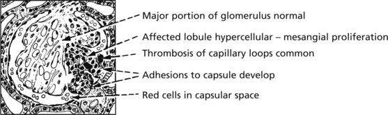
The pattern is associated with haematuria or nephrotic syndrome. Segmental lesions heal by fibrosis.
Focal Segmental Glomerulosclerosis (FSGS)
In this pattern of disease, a segment of glomerulus undergoes sclerosis without inflammation, but with an increase in mesangial matrix.
Originally regarded as a variant of minimal change nephropathy, primary FSGS presents as with haematoma or nephrotic syndrome with progression to chronic renal failure. It may recur within a transplanted kidney.
Secondary FSGS may complicate a variety of preexisting conditions including those reflux nephropathy and intravenous drug use.
IgA Nephropathy
This is the commonest form of glomerulonephritis worldwide. It can present with microscopic or macroscopic haematuria or the nephrotic syndrome and may lead to chronic renal failure. It typically affects young males, who often suffer recurrent episodes after upper respiratory infections although all ages can be affected.
The serum IgA level is raised and glomerular damage is due to IgA immune complexes. Sometimes crescentic glomerulonephritis is seen.
Mechanism
IgA immune complexes in blood → Glomeruli → Often focal deposition → Activation of complement (C3) → Often focal damage
Shows deposits of IgA and C3 within the mesangium: the capillary loops are not usually affected.
Note: The focal deposition is dependent on the molecular size of the complex and failure of mesangial clearance.
Henoch–Schönlein purpura has similar renal changes but also skin rash and gastrointestinal symptoms.
Minimal Change Glomerulonephritis
This disorder affects any age but is the major cause of nephrotic syndrome in children. It typically remits spontaneously and responds to a short course of steroids. Recurrences are quite common.
The glomeruli are histologically normal, but electron microscopy reveals fusion of podocyte foot processes. This is a finding in many causes of proteinuria. No immune complexes are found on immunofluorescence or electron microscopy.
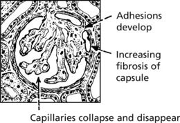
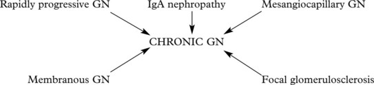
Chronic Glomerulonephritis (GN)
This is the end stage of many forms of glomerulonephritis, but most patients present at this stage without a history of previous renal disease.
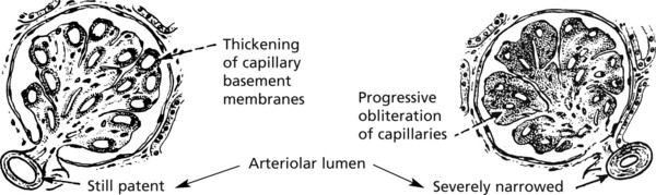
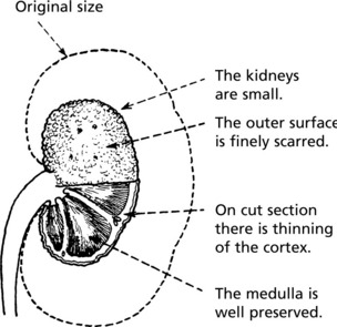
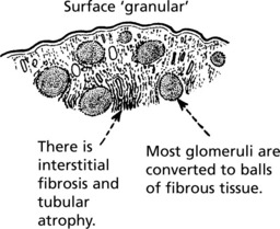
Pathology The kidneys are both small – granular contracted kidney.
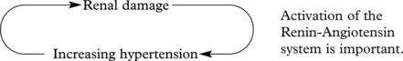
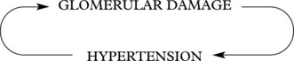
In chronic glomerulonephritis a vicious cycle is set up.
Patients may first present with chronic renal failure, or this may develop after years of glomerulonephritis.
Glomerulonephritis – Disease Mechanisms
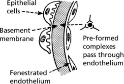
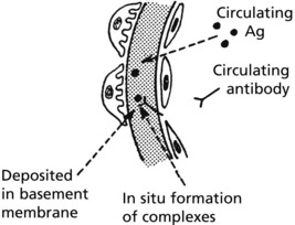
The mechanism in most forms of GN is:
The glomerulus is central to immune complex deposition and formation due to its fenestrated endothelium and high intraluminal pressure.
Immune complexes can occur in glomeruli as follows:
Localisation of Complexes
This depends on the size of the complexes, their shape and electrical charge, and their ability to penetrate the basement membrane.
Other factors modifying the pathological changes are:
Note: In addition to its damaging consequences, complement activation does solubilise complexes allowing their disposal. Thus complement deficiency may predispose to immune complex disease, e.g. in systemic lupus erythematosus (SLE).
Glomerular Disease in Systemic Disorders
Systemic Lupus Erythematosus (SLE)
At least 50% of patients with SLE have clinical evidence of renal involvement, and almost all will have abnormalities on renal biopsy.
The renal changes form a spectrum.
On immunofluorescence, IgG, C3 are almost always present and IgA, IgM, Clq and C4 are often also seen.
Amyloidosis
The general features of amyloidosis have already been described (p.24). Amyloid is deposited around the capillary basement membranes of the glomeruli and in the renal vessels and interstitium.
Gross proteinuria leading to a nephrotic syndrome is common. Interstitial fibrosis results from tubular degeneration and ischaemia due to glomerular and arteriolar lesions. Chronic renal failure results.

Full access? Get Clinical Tree

