Chapter 9 Gastrointestinal Tract
The Mouth
Viral Infections
Herpes simplex virus (usually type I) infects the mouth in young children – this is often asymptomatic but in 10–20% of cases numerous vesicles and ulcers are seen. The virus can become latent in the trigeminal ganglion and repeated ‘cold sores’ occur on the lip in later life.
Coxsackie viruses can cause oral blistering (e.g. herpangina; hand, foot and mouth disease). Koplik’s spots are a feature of measles.
Fungal Infection
Candida albicans is an oral commensal in 40% of the population.
Clinical infection is seen in infants, in patients on broad spectrum antibiotics, steroid or cytotoxic therapy and also in the immunosuppressed and diabetics. Extensive oral candida is common in patients with AIDS.
Bacterial Infections
Syphilis, now uncommon in the West, can involve the mouth as a primary chancre, irregular lines of ulceration – snail track ulcers of the secondary stage, and as small gummas (tertiary).
Oral tuberculosis, usually due to coughed up bacilli from pulmonary TB, is now very uncommon. Ulcers may occur on the tongue.
Oral Cancer
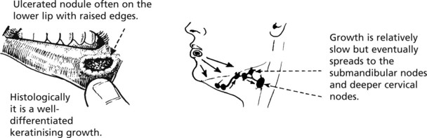
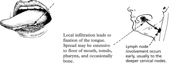
Oral cancer accounts for 2% of cancers in the United Kingdom. There is wide geographical variation – commoner in S. East Asia. Males are at least twice as commonly affected as women – it is a disease of the elderly.
Mouth
Erythroplakia and Leukoplakia
These terms describe velvety red patches and white patches in the oral mucosa. These are important because they may represent dysplasia of the squamous epithelium and may lead to squamous cancer.
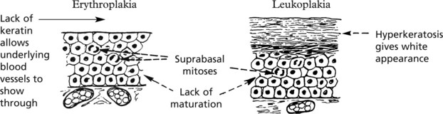
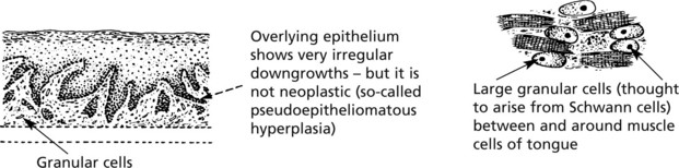
Not all examples of leukoplakia are premalignant and may also be due to:
A distinctive form – ‘hairy leukoplakia’ – occurs on the lateral border of the tongue in patients with HIV/AIDS. It is due to Epstein–Barr virus infection, often with superimposed candida.
Pigmentations
Melanotic pigmentation of the mouth is seen in Addison’s disease, haemochromatosis and the Peutz–Jeghers syndrome.
Benign Tumours
A variety of benign tumours are seen, e.g. squamous papillomas and haemangiomas (often on the lips or tongue).
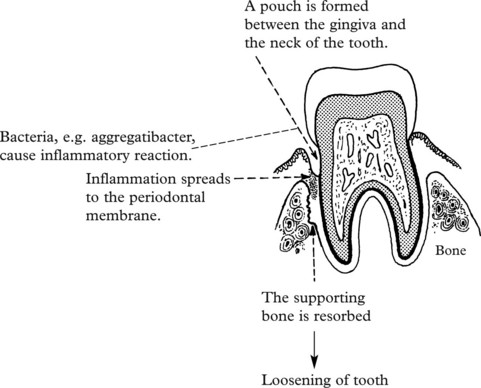
Dental Caries and Periodontal Disease
These two very common processes are primarily of importance to dentists but an understanding is also valuable for doctors.
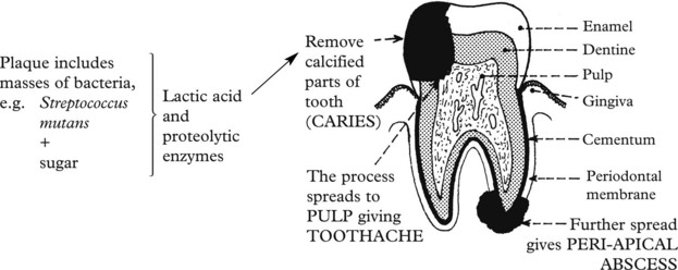
Dental Caries
This is the commonest disease of teeth.
Poor oral hygiene + high sugar intake lead to formation of plaque.
Diseases of the Salivary Glands
Inflammation
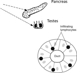
The commonest acute inflammation is due to the mumps virus, which produces acute swelling, particularly of the parotid glands, with oedema and mononuclear infiltration of the interstitial tissue.
The testes and pancreas may also be inflamed and atrophy may follow.
Bacterial infection of these glands is uncommon and may occur during a prolonged illness, particularly if a calculus has formed in a duct.
Chronic inflammation is rare. It may occur in sarcoidosis. The parotid gland becomes swollen and there may be an accompanying irido-cyclitis. This has given rise to the term ‘uveo-parotid fever’. The parotid gland shows a chronic inflammatory reaction with granulomas typical of sarcoidosis.
Sjögren’s Syndrome
In this auto-immune condition there is destruction of the salivary, lacrimal and conjunctival glands by an infiltrate of lymphocytes (so-called lympho-epithelial lesions) and plasma cells. The duct epithelia often undergo reactive hyperplasia. It results in dryness of the mouth, caries due to lack of saliva, and ulceration of the conjunctiva caused by lack of secretion from the lacrimal and conjunctival glands. This is often associated with rheumatoid disease.
Salivary Glands – Benign Tumours
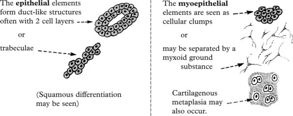
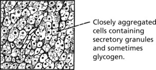
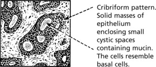
Pleomorphic Adenoma
This is the commonest tumour of the salivary glands and most often occurs in the parotid. The term ‘pleomorphic’ applies not to the nuclei of the cells but to the different types of tissue found. These are derived from the epithelial and myoepithelial cells.
The tumour is lobulated and encapsulated but there are frequently small lobules which extend into the adjacent tissue.
If the tumour is ‘shelled out’ these lobules are left behind and cause local recurrence. For this reason superficial parotidectomy is usually performed, ensuring complete removal of the tumour.
Malignant Change
Under 5% of these tumours become malignant, often after many years. Most of these are adenocarcinomas with a poor prognosis.
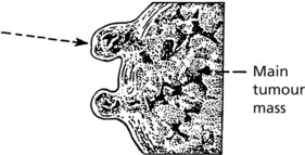
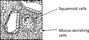
Salivary Glands – Carcinomas
Almost all malignant tumours of the salivary glands are adenocarcinomas. They affect major and minor glands and arise de novo or from pre-existing pleomorphic adenoma. The prognosis is variable.
Three unusual subtypes are worth noting.
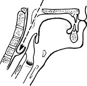
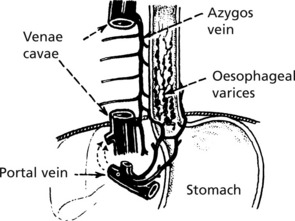
Approximately 1/4 of all malignant parotid tumours are of this type. It grows slowly, recurs after removal and sometimes there is late spread to the regional lymph nodes and distant organs.
This usually occurs in minor salivary glands. It extends by direct spread, especially along nerve sheaths, but metastases can occur to lymph nodes, lungs and bones.
Oesophagus
The oesophagus is a muscular tube 25 cm long lined by stratified squamous epithelium which is resistant to damage by heat, cold and mechanical trauma.
Inflammation
Reflux Oesophagitis
This is the commonest form of inflammation due to reflux of gastric acid through a relaxed lower oesophageal sphincter into the lower oesophagus, often associated with hiatus hernia.
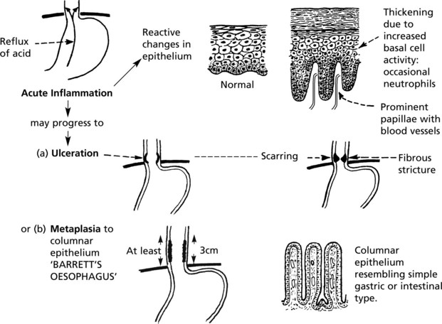
This is an important premalignant lesion with a 30–40 fold increased risk of cancer.
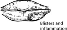
Other Forms of Oesophagitis
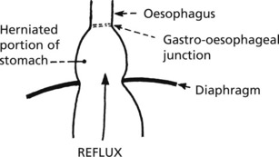
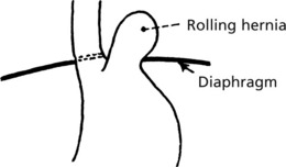
Hiatus Hernia
In hiatus hernia part of the stomach herniates into the thorax. This is common, particularly in the elderly, though often asymptomatic. Two forms are seen:
Diverticula
These are relatively rare and are of 2 varieties.
Involves pharynx (pharyngeal pouch). Sac is distended during swallowing of food. By pushing down behind oesophagus, it may compress this structure. Abnormal function of the upper oesophageal sphincter is an aetiological factor.
This is due to traction of fibrous tissue produced by mediastinal inflammation, e.g. tuberculosis of lymph nodes.
Rarely, there may be a congenital diverticulum at the level of the bifurcation of the trachea.
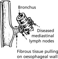
Obstruction usually leads to dysphagia – difficulty in swallowing. The causes include:
Strictures of the oesophageal wall due to:
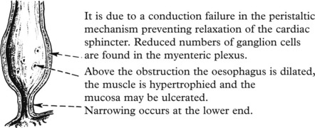
Achalasia of the oesophagus develops in young adults and may cause severe obstruction.
(In South American trypanosomiasis the myenteric plexus may be destroyed (CHAGAS DISEASE) and long-standing diabetic autonomic neuropathy may cause a similar problem.)
In Systemic sclerosis, replacement of muscle by fibrous tissue converts the oesophagus into a rigid tube.
Oesophageal Varices
These dilated veins occur secondarily to portal hypertension caused mainly by cirrhosis of the liver (see p.350).
Fibrosis in the liver obstructs the flow of blood from the gastrointestinal tract. Anastomotic channels connecting the portal and systemic venous systems open up and become distended. The most important are the oesophageal tributaries of the azygos vein which connect through the diaphragm with the portal system. They become varicose, and are easily traumatised by the passage of food, leading to haemorrhage which can be severe.
Spontaneous rupture of the oesophagus is rare. Mucosal tears causing haemorrhage may occur (Mallory–Weiss syndrome).
Congenital abnormalities include stenosis and atresia with fistula formation.
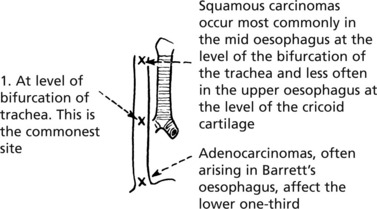
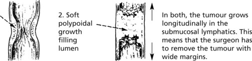
Oesophagus – Tumours
Benign tumours of the oesophagus are rare. They are almost always of connective tissue origin (usually leiomyomas) and form polyps within the lumen, causing obstruction.
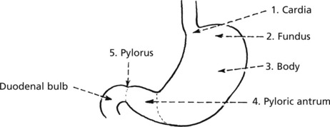
Carcinoma
Carcinoma of the oesophagus occurs in two main forms:
The disease is more common in men by at least 4 to 1.
The tumours may narrow the lumen or cause a polypoid mass
Spread
Middle third tumours may involve the trachea with fistula formation leading to aspiration pneumonia. Tumours of the lower third may invade the mediastinum. Lymph node involvement is common and blood borne metastases to liver occur late.
Aetiology
There is geographical variation, tumours being common in China and Africa. Smoking and diet, including alcohol consumption, are important in squamous carcinoma. Post-cricoid carcinoma in women is a rare late complication of the dysphagia complicating iron deficiency anaemia. Adenocarcinoma is largely due to Barrett’s oesophagus and reflux.
Stomach
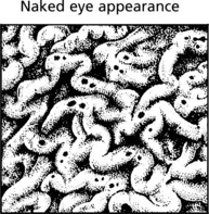
The stomach is divided into five anatomical regions:
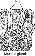
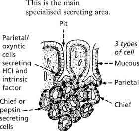
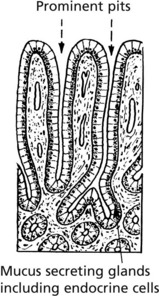
Three forms of mucosa are seen:
Acute Gastritis
Mild acute gastritis with neutrophils in the mucosa may be caused by alcohol and non-steroidal anti-inflammatory drugs (NSAIDs), and are seldom biopsied. Acute haemorrhagic or erosive gastritis is a more severe form, also associated with aspirin and NSAIDs, and is also a complication of shock.
Tiny ulcers affect all parts of the stomach, occuring on the apex of mucosal folds, and can heal rapidly.
Gastritis
Acute Inflammation
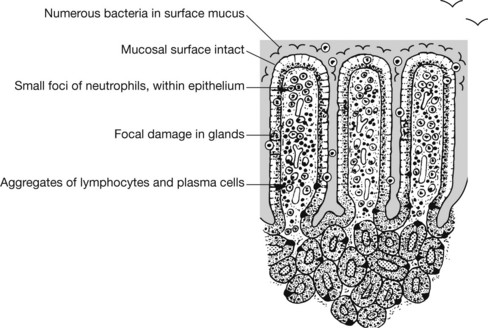
Mild acute gastritis is an acute inflammation with neutrophil reaction in the superficial layers of the mucosa. Pain and sickness have a multitude of causes varying from hot fluids, alcohol and aspirin which act as direct irritants, to infections such as childhood fevers, viral infections and bacterial food poisoning.
A more acute form, known as acute haemorrhagic or erosive gastritis, is associated with ingestion of irritant drugs, particularly aspirin and NSAIDs and is also a complication of shock states.
Haemorrhagic Erosions
These are tiny ulcers, a few millimetres in diameter, which are formed by the digestion of the mucous membrane overlying small haemorrhages. They are usually multiple and affect all parts of the stomach. They occur mostly on the apex of mucosal folds and involve only the mucosa.
Note that the changes are superficial so that restoration to normal can occur very quickly occur.
Chronic Gastritis
There are 3 main causes of chronic gastritis:

Full access? Get Clinical Tree

