Gastric Antral Vascular Ectasia
Amitabh Srivastava, MD
Key Facts
Terminology
GAVE, “watermelon stomach”
Etiology/Pathogenesis
Elderly patients, often with associated connective tissue disease
Clinical Issues
Upper GI bleeding or chronic occult blood loss
Endoscopic laser coagulation therapy is treatment of choice
Macroscopic Features
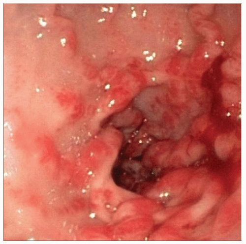
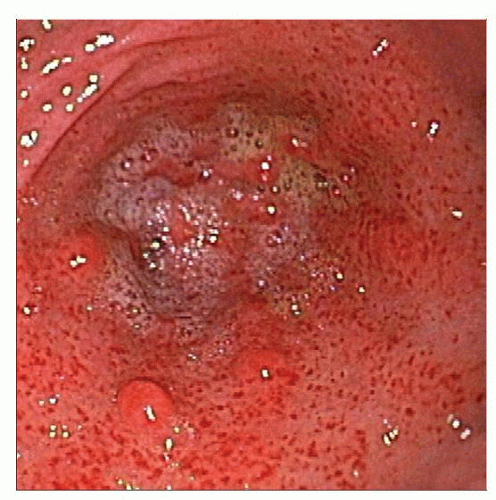
Raised red mucosal stripes converging on pylorus (“watermelon stomach”)
Microscopic Pathology
Vascular ectasia, fibrin thrombi, and myofibroblastic proliferation in antral biopsies
 Endoscopic photograph shows typical findings in gastric antral vascular ectasia, including raised linear red stripes converging on the pylorus (“watermelon stomach”). |
TERMINOLOGY
Abbreviations
Gastric antral vascular ectasia (GAVE)
Synonyms
“Watermelon stomach”
Definitions
Vascular lesion involving gastric antrum
Combination of vascular ectasia, fibrin thrombi, and myofibroblastic proliferation
ETIOLOGY/PATHOGENESIS
Postulated Causes
Degenerative changes
Abnormal antral motility, mechanical stress, and mucosal prolapse
Endogenous peptide = mediated vascular dilatation
Serum gastrin or prostaglandin E2-mediated vascular ectasia
Portal hypertension does not play direct role in pathogenesis
CLINICAL ISSUES
Epidemiology
Incidence
Exact incidence unknown (only symptomatic patients undergo examination)
4% of nonvariceal upper GI bleeding estimated to be related to GAVE
Age
Typically in elderly patients
Gender
Associated with autoimmune conditions in older women (mean age > 70 years)
Cirrhosis typically seen in men who are slightly younger (mean age = 65 years)


