As discussed earlier, normal meiosis requires pairing of homologous chromosomes followed by recombination. The autosomes and the X chromosomes in females present no unusual difficulties in this regard; but what of the X and Y chromosomes during spermatogenesis? Although the X and Y chromosomes are different and are not homologues in a strict sense, they do have relatively short identical segments at the ends of their respective short arms (Xp and Yp) and long arms (Xq and Yq) (see Chapter 6). Pairing and crossing over occurs in both regions during meiosis I. These homologous segments are called pseudoautosomal to reflect their autosome-like pairing and recombination behavior, despite being on different sex chromosomes.
Oogenesis
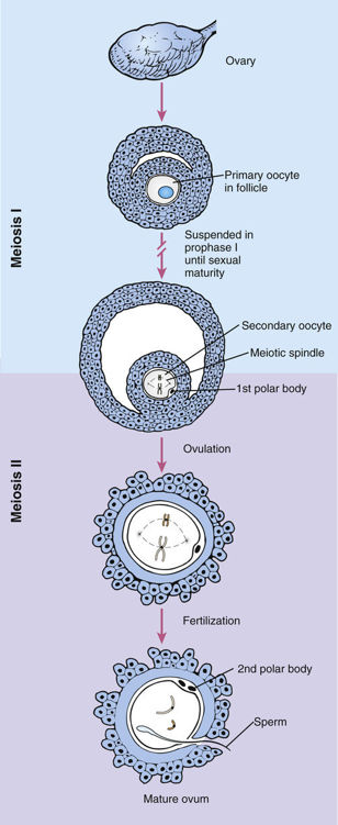
Whereas spermatogenesis is initiated only at the time of puberty, oogenesis begins during a female’s development as a fetus (Fig. 2-17). The ova develop from oogonia, cells in the ovarian cortex that have descended from the primordial germ cells by a series of approximately 20 mitoses. Each oogonium is the central cell in a developing follicle. By approximately the third month of fetal development, the oogonia of the embryo have begun to develop into primary oocytes, most of which have already entered prophase of meiosis I. The process of oogenesis is not synchronized, and both early and late stages coexist in the fetal ovary. Although there are several million oocytes at the time of birth, most of these degenerate; the others remain arrested in prophase I (see Fig. 2-14) for decades. Only approximately 400 eventually mature and are ovulated as part of a woman’s menstrual cycle.


Stay updated, free articles. Join our Telegram channel

Full access? Get Clinical Tree


