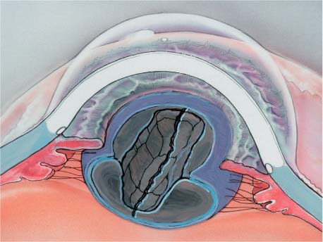
Chapter 10 Attention to surgical detail and knowledge of alternative techniques are especially important in performing a complication-free phacoemulsification procedure. This chapter describes the major phases of the four-quadrant in situ phacoemulsification technique,1 commonly called “divide and conquer,” the flow of the procedure, typical problem-causing areas, and solutions to common problems. Specific details of each original surgical technique, variations, and applications are found in the references and in appropriate courses. Nuclear splitting techniques1–3 evolved and were popularized following the advent of capsulorrhexis,4 which made subluxation of the lens nucleus into the pupillary plane difficult. These techniques enabled the surgeon to confine emulsification within the capsular bag and more easily manage smaller sections of the nucleus. The nucleus may be divided into sections or quadrants with vertical splits1,2 or delaminated internally by circumferentially separating an inner from an outer nucleus5; the two approaches may be combined by delaminating the nucleus and then splitting the internucleus vertically.3 Figure 10–1 shows the locations of lens nucleus areas created by the phacoemulsification sculpting process that will be referred to in this chapter. If there is a tear in the edge of the anterior capsulotomy, some of the maneuvers required for this nucleus management technique, such as nucleus rotation and cracking, might cause the tear to extend peripherally and then posteriorly. If the capsulotomy edge is torn after the nucleus has been hydrodissected and freely rotated, cracking may be safely accomplished, provided the crack is oriented perpendicular to, rather than directed toward, the capsular tear. If the nucleus has not been completely loosened by hydrodissection, it will be difficult or impossible to rotate. Often this is not appreciated until after the first trough has been sculpted and an attempt is made to rotate the nucleus to sculpt the second trough. In this situation the procedure should be interrupted so that repeat hydrodissection of the nucleus can be accomplished. This will allow for free rotation of the nucleus prior to continuing with the emulsification procedure. Failure to do so risks compromising zonular integrity. FIGURE 10–1 Illustration of a sculpted trough, trough wall, residual posterior lens/cortical plate, and a crack in the posterior lens/cortical plate. The consistency of the lens nucleus will dictate which emulsification techniques can be most successfully employed. The four-quadrant divide and conquer technique fortunately has a very broad application. The surgeon may encounter difficulty only with lenses that are very soft or very hard. If the lens consistency is very soft, the surgeon will initially note little resistance to sculpting during the creation of the first trough. If the lens material seems to “jump” toward the aspiration port more than would be expected, the lens is soft, that is, more cortical in nature. The surgeon, therefore, may encounter difficulty when cracking is attempted. The instruments used may seem to cut through the walls of the trough and the lens material like a wire cheese cutter through cheese, rather than spreading the walls of the trough, thus creating a crack in the posterior lens plate. If the lens is recognized to be softer than expected during the sculpting of the initial trough, shifting to an alternative technique, such as dividing the lens into two heminuclei and subluxing them into the anterior chamber utilizing hydrodissection and then removal using aspiration and low-power phaco, should be considered. In some cases the softness of the lens will not have been appreciated during the sculpting maneuver, but when cracking is attempted the walls of the trough crumble rather than spread apart, preventing a crack from taking place. This friable type of lens will still permit the divide-and-conquer technique provided the trough is deep enough, thus minimizing the amount of posterior plate that must be cracked. Cessation of the cracking maneuver to re-deepen the trough before trying to complete the cracking is appropriate. Lenses that are very hard may present a challenge not only during the sculpting of the troughs but also during the cracking of the posterior plate. Hard lenses require an increase in ultrasonic power and a decrease in the depth of each emulsification pass across the lens so that the lens does not appear to be pushed away from the ultrasound tip during the sculpting process. They also require sculpting farther out into the periphery. If the ultrasound tip appears to be pushing the lens during the sculpting pass, the phaco power should be increased. An alternative tip that maximizes cavitation power, such as an angled Kelman tip, may be employed. Converting to an extracapsular technique may be indicated. Often, after increasing the ultrasonic power sufficiently to make progress with emulsification, an increase in cavitation bubbles collecting in the anterior chamber will occur. These bubbles will obstruct the view, and should prompt consideration of whether or not it is prudent to continue with this technique in view of the increased time and power that will be required to complete lens removal. Harder lenses will be more difficult to split unless the trough sculpted in the lenses is deep, leaving a very thin nuclear/cortical plate. Deep sculpting in a hard lens is a time-intensive effort. Low vacuum settings (dependent on phaco machine capability) help prevent high-power encounters with the iris, as well as aspiration of the equatorial bag, during sculpting. If the hard lens is recognized preoperatively, a large capsulotomy should be made to retain the ability to change to an alternative lens management technique and avoid becoming “imprisoned” in the capsular bag by too small a capsulotomy opening. To split the nucleus, especially if it is hard (nuclear mature), the splitting instruments must be placed at the base of the trough. This will place the vectors for splitting the nucleus in the proper orientation to create a split (Fig. 10–2A). If the instruments are too high in the trough, the posterior plate will be bent but not split (Fig. 10–2B).
FOUR-QUADRANT DIVIDE
AND CONQUER
HISTORY
PHACOEMULSIFICATION STAGES AND PROBLEMS ENCOUNTERED
CAPSULOTOMY NONCONTIGUOUS
INCOMPLETE HYDRODISSECTION
LENS CONSISTENCY
Soft Lens
Hard Lens
Stay updated, free articles. Join our Telegram channel

Full access? Get Clinical Tree


