Visual field deficits.
What is the role of ophthalmoscopy in patients presenting with headache?
Ophthalmoscopy serves to identify:
How do you isolate the contributions of the individual muscles when testing eye movements?
On confrontation, ask the patient to fix his or her gaze on your finger while you draw a standard ‘H’ pattern in the air. If the patient is not able to keep the head still, use your free hand to stabilise the chin. The H-shape allows testing of vertical eye movements in abduction and adduction, to which the muscles contribute as follows:
| Muscle | Nerve | Action | |
|---|---|---|---|
| Medial rectus | III | Adduction | |
| Lateral rectus | VI | Abduction | |
| (In adduction) | (In abduction) | ||
| Superior rectus | III | (Intorsion) | Elevation |
| Inferior rectus | III | (Extorsion) | Depression |
| Superior oblique | IV | Depression | (Intorsion) |
| Inferior oblique | III | Elevation | (Extorsion) |
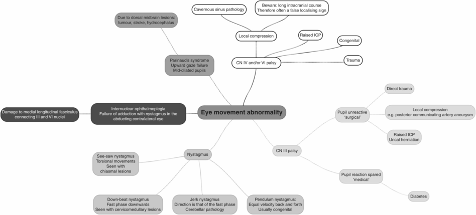
What are the characteristics of a cranial nerve III palsy?
The patient suffers diplopia, which may be reduced on abduction of the affected eye. The eye is deviated ‘down and out’ at rest. This is associated with ptosis and mydriasis (pupil dilatation).
How would you distinguish the divisions of the trigeminal nerve on examination?
The divisions are tested using light touch stimulus:
Va (ophthalmic): on the forehead
Vb (maxillary): over cheekbone
Vc (mandibular): on the mandible
Only Vc conveys a motor component, supplying the muscles of mastication.
How would you distinguish upper and lower motor neuron pathology in the facial nerve?
In upper motor neuron lesions the movement in the upper part of the face is spared because of bilateral supranuclear inputs to the nuclei controlling the frontalis and orbicularis occuli muscles.
Mild weakness may still manifest as an inability to ‘bury’ the eyelashes on forced eye closure.
What is Bell’s phenomenon?
Bell’s phenomenon is the upward rolling of the eye observed as the eyelid fails to close – this serves to protect the cornea when eye closure is impaired, typically due to lower motor neuron damage to the facial nerve.
How would you perform Weber’s and Rinne’s tests?
Weber’s test (useful screening test): place a resonating tuning fork in the centre of the patient’s forehead. The sound should be heard equally on each side.
To differentiate between the two perform Rinne’s test.
Rinne’s test: place a resonating tuning fork on the mastoid process. Once sound can no longer be detected by the patient through the mastoid, place the tuning fork adjacent to the ear canal to determine whether residual vibrations are still heard. This compares bone and air conduction, respectively.
To exclude a conductive hearing loss – air conduction should be louder than bone conduction.
For further details see Chapter 32, Examination of the ear.
How would you assess vestibular function?
Vestibular function can be assessed by:
How would you assess cranial nerves IX and X?
The gag reflex is subserved by the glossopharyngeal nerve (CN IX) as the afferent limb and the vagus nerve (CN X) as the efferent limb. However, it is unpleasant for the subject, so observing the patient’s cough and swallow is the more appropriate routine screen in conscious patients.
What are you testing by asking the patient to turn the head to the right?
The left sternocleidomastoid muscle (CN XI) turns the head to the right, and the right SCM turns it to the left.
In which direction does the tongue deviate in the context of a CN XII lesion?
The tongue deviates to the side of the lesion on protrusion.
Examination of the peripheral nervous system: upper limbs
Checklist
WIPER and Physiological parameters
Adequate exposure of neck and arms
Inspection
Neck and arms:
wasting, fasciculation, contractures
scars over the cervical spine
skin stigmata (neurofibromas)
Pronator drift
Tone
Wrist, elbow and shoulder assessed in flexion and extension
Normal/increased/decreased
Power
Grade MRC 1–5 in all groups
Coordination
Finger–nose
Reflexes
Biceps, triceps, supinator
Hoffmann’s sign
Sensation
Fine touch
Joint position (proprioception)
Examination notes: upper limbs
What cervical spine scars should be identified?
Posterior scars:
decompression, laminectomy
foramen magnum decompression (for Chiari malformation)
Anterior scars:
decompression/discectomy
How is muscle power graded?
Stay updated, free articles. Join our Telegram channel

Full access? Get Clinical Tree


