Fig. 29.1
Body fluid compartments in terms of volumes of fluid
The ECF can be further subdivided into:
Interstitial fluid compartment, water in dense connective tissue and water of bone and transcellular fluid compartment (ISF) – 75 % of ECF
Intravascular fluid compartment (Plasma) – 25 % of ECF
The transcellular fluid compartment consists of a collection of fluids, formed during the process of the transport activities in cells and is found mainly in epithelial lined spaces. They are important because of the special roles they play in the body. Examples of transcellular fluids are cerebrospinal fluid, joint fluid, fluid in the bowel, fluid in body cavities, aqueous humor, bile in the gall bladder and urine in the bladder.
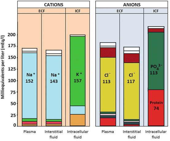
All of these fluids contain electrolytes (ions) that are distributed in such a way as to maintain electrical neutrality (Fig. 29.2). Potassium is the most abundant electrolyte in the ICF and sodium in the ECF. The difference is maintained by the basolateral Na+/K+ ATPases, which transport three Na+ ions out of the cell in exchange for two K+ ions transported into the cell. A balance of charges is maintained in each compartment by different ions.


Fig. 29.2
Electrolyte concentrations in ECF and ICF
Major fluid and electrolyte shifts occur daily between the various fluid compartments. Some of these fluid shifts require energy, others do not. In the acutely ill patients, these shifts are even more exaggerated.
Water moves passively across semi-permeable membranes from areas of lower to higher osmolality by a process called osmosis. Osmolality refers to the concentration of osmotically active particles, like proteins, expressed in terms of osmoles of solutes per kilogram of solvent. The ICF is hypertonic, or hyperosmolar, compared to the interstitial fluid, so water will move into the cell by osmosis. This will occur until the osmolality is the same on both sides of the membrane. This may explain the higher proportion of intracellular water compared to extra-cellular water (28 vs 14 L in a 70-kg man). If the osmolality of the ECF were increased (as in dehydration), then there would be net water movement out of cells and this would continue until the osmolality in the ICF equals that of the ECF. However, as the osmotic pressure between the intracellular compartment and the interstitial space is minimal, there is very little fluid movement between these two areas.
In normal healthy cells, active transport maintains a high concentration of potassium and phosphate and a low concentration of sodium within the cell. There is thus a constant tendency for these electrolytes (i.e., potassium and phosphate) to diffuse down the concentration gradient out from the cell. The sodium-potassium pump, in an energy-requiring process, constantly pumps the potassium back into the cell in exchange for sodium, maintaining the electrolyte concentrations, resulting in a potential difference across the membranes of the cells, which is vital for cell function and life. As long as there is enough energy to maintain the function of the sodium-potassium pumps, the ionic distribution is maintained and there is homeostasis.
At the level of the capillary, oxygen, ions and nutrients (in the form of electrolytes, glucose, amino acids and lipids) pass from the blood to the interstitial fluid compartment where they subsequently move into the cells. Waste products (carbon dioxide and acids) from the cells move in the opposite direction from the cells back to the capillaries through the interstitial space.
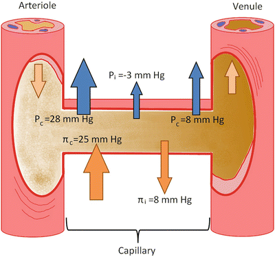
Fluid movement at this site is determined by the differences in hydrostatic and oncotic pressures between the capillaries and interstitial fluid. These forces, also known as Starling forces, result in net movement of fluid into or out of the capillaries (Fig. 29.3). The normal fluid transfer occurs out of the capillaries and amounts to about 1 ml per 100 g tissue per minute.


Fig. 29.3
Starling forces across the capillary. The Starling forces (hydrostatic [Pc and Pi in the capillary and interstitium respectively] and oncotic pressures [πc and πi in capillary and interstitium respectively]) allow for bulk flow of fluid and nutrients across the capillary wall
The principal determinants of transvascular exchange, has always been thought to be due to the Starling forces across the capillaries. Recent findings have questioned these classic models. Non-fenestrated (continuous) capillaries (see Chap. 4, Fig. 4.2) throughout the interstitial space have been identified, indicating that absorption of fluid does not occur through venous capillaries but that fluid from the interstitial space, which enters through a small number of large pores, is returned to the circulation primarily as lymph that is regulated through sympathetically mediated responses.
Stay updated, free articles. Join our Telegram channel

Full access? Get Clinical Tree


