Fibroma of Tendon Sheath
David R. Lucas, MD
Key Facts
Terminology
Small benign fibrous nodule typically attached to tendon sheath
Clinical Issues
Most common in fingers
Macroscopic Features
Median size: ≈ 2 cm (range: 0.5-5 cm)
Microscopic Pathology
Well demarcated
Attached to tendon or tendon sheath
Benign fibroblasts and myofibroblasts
Slit-like vascular spaces
Cellular nodular fasciitis-like areas
Top Differential Diagnoses
Nodular fasciitis
Superficial fibromatosis
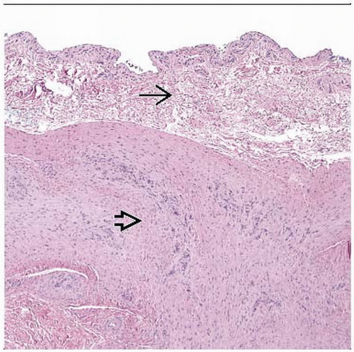 Fibroma of tendon sheath (FTS) typically presents as a small soft tissue nodule in a finger. This micrograph depicts a circumscribed fibroma  attached to a synovial-lined tendon sheath attached to a synovial-lined tendon sheath  . . |
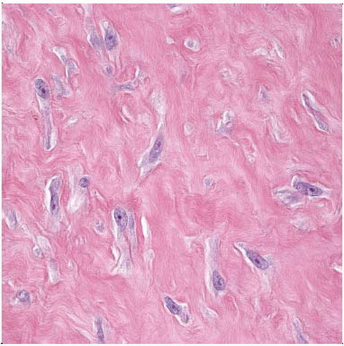 The proliferating cells in FTS are fibroblasts and myofibroblasts characterized by bipolar spindle and stellate cells with abundant amphophilic cytoplasm and vesicular nuclei with solitary nucleoli. |
TERMINOLOGY
Abbreviations
Fibroma of tendon sheath (FTS)
Definitions
Small benign fibrous nodule typically attached to tendon sheath
ETIOLOGY/PATHOGENESIS
Histogenesis
Generally regarded as reactive nonneoplastic process
Single report of clonal chromosomal aberration t(2:11)
Possibly neoplastic process
CLINICAL ISSUES
Epidemiology
Incidence
Uncommon, exact incidence unknown
Age
Stay updated, free articles. Join our Telegram channel

Full access? Get Clinical Tree


