Extranodal NK/T-cell Lymphoma, Nasal Type
Aaron Auerbach, MD, PhD
Key Facts
Terminology
Tumor of NK cells or T cells, with necrosis, Epstein-Barr virus infection, and angioinvasion
Etiology/Pathogenesis
Epstein-Barr virus
Clinical Issues
Common in adults in South America, Central America, and Asia
Nasopharynx most common site, skin is 2nd
Microscopic Pathology
Diffuse dermal/subcutaneous infiltrate of variably sized tumor cells, angiocentric with necrosis
Dense chromatin, can be vesicular in large cells
Coagulative necrosis in most cases
Ancillary Tests
Epstein-Barr virus antigen (+)
Mostly NK-cell markers phenotype
βF1(−), CD2(+), cytoplasmic CD3-ε(+), CD56(+/-), cytotoxic markers(+)
Less often T-cell marker phenotype
βF1(+), CD2(+), cytoplasmic CD3-ε(+), CD56(-/+), cytotoxic markers(+)
No specific cytogenetic abnormalities
Top Differential Diagnoses
Wegener granulomatosis
Infection
B-cell lymphomas (DLBCL, lymphomatoid granulomatosis)
PTCL-NOS, NK-cell leukemia
TERMINOLOGY
Abbreviations
Extranodal NK/T-cell lymphoma, nasal type (ENNKTCL)
Synonyms
Angiocentric T-cell lymphoma, malignant midline reticulosis, lethal midline granuloma, polymorphic reticulosis
Definitions
Lymphoma composed of NK cells or T cells that is characterized by necrosis, Epstein-Barr virus infection, and often angioinvasion
Usually extranodal
ETIOLOGY/PATHOGENESIS
Infectious Agents
Epstein-Barr virus
Epstein-Barr virus antigens are identified by immunohistochemistry and in situ hybridization
Immunosuppression
Subset of patients are immunosuppressed
Including post transplant
CLINICAL ISSUES
Epidemiology
Incidence
Rare in United States; more common in South America, Central America, and Asia
Age
Usually adults
Site
Nasal
Most common location of disease
Anywhere in upper aerodigestive tract
Includes nasal cavity, nasopharynx, sinuses, and palate
Extranasal
Skin
Most common extranasal site
Skin can be primary disease or secondary to disease elsewhere
Often extremities or torso
Other sites include gastrointestinal tract, lymph nodes, and testes
Presentation
Skin
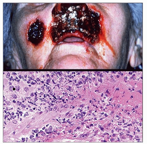
Nodules &/or plaques on skin
Often ulcerated
Nasal
Obstruction or epistaxis
Gastrointestinal tract
Perforation or mass
B symptoms including fever &/or weight loss
Hemophagocytic syndrome in some cases Laboratory Tests
EBV DNA can be assessed to check disease activity
Natural History
Can spread to regional lymph nodes
Tumor only in lymph nodes and not other anatomic sites is uncommon
Primary lymph node involvement is very rare
Can involve bone marrow
Treatment
Chemotherapy and radiation therapy Prognosis
Median survival < 15 months
Better survival with intense radiotherapy and chemotherapy
Poor prognostic indicators
High stage of disease
Bone marrow with EBV(+) cells
High International or Korean Prognostic Index
B symptoms, serum LDH, regional lymph nodes, stage
↑ EBV DNA
↑ C-reactive protein, thrombocytopenia
↑ Ki-67 > 50%
Better prognostic indicators
More favorable for nasal disease than extra-nasal
High absolute lymphocyte count
CD56(+), CD30(+) coexpression has been reported
IMAGE FINDINGS
General Features
Mass lesion that can destroy bone, especially in nasal tumors
MACROSCOPIC FEATURES
General Features
1 or more firm nodules
MICROSCOPIC PATHOLOGY
Histologic Features
Skin/mucosa
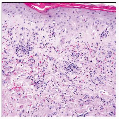
Diffuse pattern of infiltration
Mostly dermal infiltrate, sometimes subcutis
Rare foci of epidermotropism in 30%
Tumor cells can be small, medium, or large
Irregular nuclear shapes
Dense chromatin, can be vesicular in large cells
Sometimes ↑ clear cytoplasm
Coagulative necrosis in most cases
Tumor cells are angiocentric and angiodestructive
Overlying epithelium/mucosa
± ulceration
Often shows pseudoepitheliomatous hyperplasia
↑ mitoses
Erythrophagocytosis is sometimes seen
Lymph node
Tumor tends to involve paracortical areas
Bone marrow
Usually interstitial infiltrate
Lacking large tumor aggregates
Involved in 15% of cases
Cytologic Features
Rarely, diagnosis can be made by fine needle aspirate
Azurophilic granules in some tumor cells
ANCILLARY TESTS
In Situ Hybridization
In situ hybridization for EBV small-encoded RNA (EBER)
Immunohistochemistry
Epstein-Barr virus antigens positive
Immunohistochemistry for latent membrane protein (LMP)
LMP is less sensitive than EBER
Stay updated, free articles. Join our Telegram channel

Full access? Get Clinical Tree


