Left posterolateral thoracotomy.
Posterolateral thoracotomy (5th intercostal space) to access the lungs and pleura
Anterolateral thoracotomy (5th intercostal space) to access heart and pleural cavity
Clamshell (bilateral 5th intercostal spaces) for trauma or transplantation
2 cm transverse incision above sternal notch for mediastinoscopy for lung cancer staging
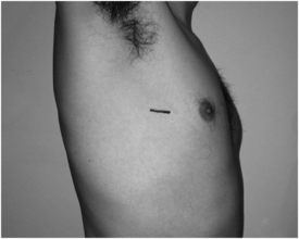
Video-assisted thoracoscopic surgery (VATS):
3 cm incision in 3rd intercostal spaces in the anterior axillary line with two or three other 1 cm incisions in 6th intercostal spaces
one to four ports are typically used, depending on procedure and pathology
Chest drains (5th intercostal space anterior axillary line)
Tattoos for radiotherapy
What are the basic checks for a patient with a chest drain?
1. What is being drained?
Establish the need for chest drainage and the nature of the effluent:
air (pneumothorax) via a flutter valve or chest drain bottle with a water seal
fluid (effusion)/pus (empyema)/blood (haemothorax) into a stoma bag or chest drain bottle with a water seal
2. Is the drain on suction?
Establish if the drain is on suction as well as the pressure applied (routine suction pressure –5 kPa).
Movement of fluid in the drain on inspiration/expiration suggests that the drain is in continuity with the thoracic cavity and is therefore working.
Stay updated, free articles. Join our Telegram channel

Full access? Get Clinical Tree


