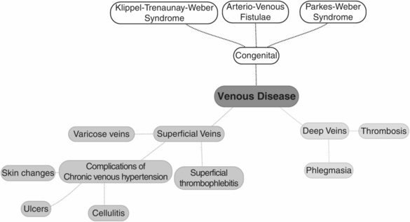
Lower limb venous anatomy.
What are the symptoms of chronic venous disease?
Leg ache, heaviness, tightness or bursting pain associated with prolonged periods of standing
Skin irritation or itchiness
Leg swelling
What is the CEAP classification?
The CEAP classification is a global method of describing the burden of venous disease, based on:
Clinical presentation (e.g. reticular veins, oedema, ulcers)
Aetiology (e.g. primary vs. secondary)
Anatomy (e.g. superifical, perforator, deep venous involvement)
Pathophysiology (e.g. reflux vs. obstruction)
What are the main perforator/communicating veins called?
Thigh – Dodd
Gastrocnemius – Boyd
Calf – Cockett
Ankle – Kuster or May
Why should the arterial system be assessed in patients with varicose veins?
Compression therapy for varicose veins can only be used in patients without peripheral arterial disease.
Any planned surgery for varicose veins in the lower limb cannot be performed in the presence of significant arterial disease, because of the risk of wound non-healing.
Always check foot pulses and ABPI.
What is phlegmasia caerulea dolens?
Phlegmasia caerulea dolens is a tender, blue discolouration of the entire limb. This is caused by acute massive venous thrombosis and obstruction of the entire venous system of the limb. It may extend proximally into the iliofemoral veins. In the short term it can lead to limb-threatening venous gangrene (ischaemia) and life-threatening pulmonary embolism. In the long term it can cause post-thrombotic limb syndrome due to venous valve damage and deep venous incompetence.
What nerve travels with the long saphenous vein?
The saphenous nerve.
What nerve travels with the short saphenous vein?
The sural nerve.
How do you inspect for varicose veins?
The patient must always be standing, preferably on an examination stool/step. The inspection has four main end points:
Has there been previous surgery for varicose veins? SFJ ligation is often performed through a groin-crease incision, which can be easily missed. Ensure you expose the groin adequately and stretch the groin-crease skin open with two fingers.
Are there any varicose veins? Determine their anatomical distribution and the vein from which they arise. Inspect all aspects of the leg by asking the patient to turn 360°.
Are there any skin changes caused by chronic venous hypertension?
Is there any evidence of deep venous hypertension (thrombosis or reflux)?
Where is the saphenofemoral junction (SFJ) located?
Palpate the pubic tubercle. The SFJ is found 2–4 cm (2 fingers’ breadths) inferior and 2–4 cm lateral to the pubic tubercle.
Stay updated, free articles. Join our Telegram channel

Full access? Get Clinical Tree


